International Orthopaedics ( IF 2.7 ) Pub Date : 2022-08-22 , DOI: 10.1007/s00264-022-05552-5 Steffen Brodt 1 , Vincent Boersch 2 , Patrick Strube 1 , Georgi Wassilew 3 , Georg Matziolis 1
|
Purpose
When revising acetabular cups, it is often necessary to provide additional stabilisation with screws. In extensive defect situations, the placement of screws caudally in the ischium and/or pubis is biomechanically advantageous. Especially after multiple revision operations, the surgeon is confronted with a reduced bone stock and unclear or altered anatomy. In addition, screw placement caudally is associated with greater risk. Therefore, the present study aims to identify and define safe zones for the placement of caudal acetabular screws.
Methods
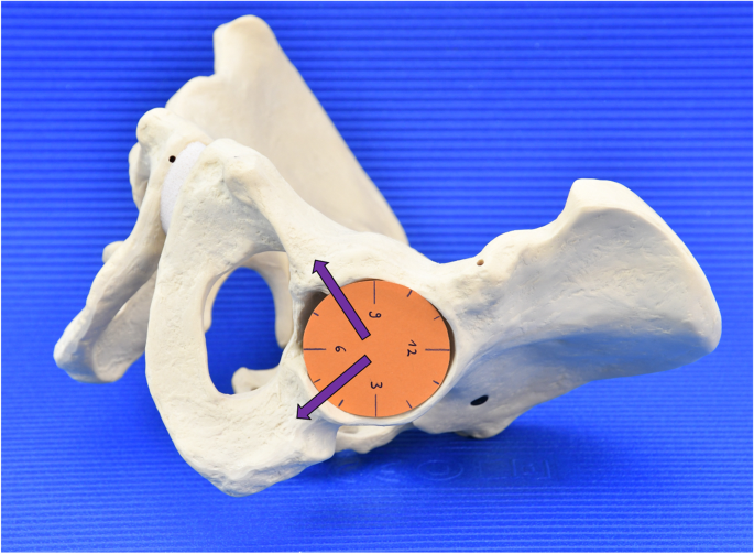
Forty-three complete CT datasets were used for the evaluation. Sixty-three distinctive 3D points representing bone landmark of interests were defined. The coordinates of these points were then used to calculate all the parameters. For simplified visualisation and intra-operative reproducibility, an analogue clock was used, with 12 o’clock indicating cranial and 6 o’clock caudal.
Results
A consistent accumulation was found at around 4.5 ± 0.3 hours for the ischium and 7.9 ± 0.3 hours for the pubic bone.
Conclusions
The anatomy of the ischium and pubis is sufficiently constant to allow the positioning of screws in a standardised way. The interindividual variation is low — regardless of gender — so that the values determined can be used to position screws safely in the ischium and pubis. The values determined can provide the surgeon with additional orientation intra-operatively when placing caudal acetabular screws.
中文翻译:
在翻修手术中定义坐骨和耻骨螺钉的管
目的
在修改髋臼杯时,通常需要使用螺钉提供额外的稳定性。在广泛的缺损情况下,将螺钉在坐骨和/或耻骨尾部放置在生物力学上是有利的。特别是在多次翻修手术后,外科医生面临着骨量减少和解剖结构不清晰或改变的问题。此外,尾部螺钉置入与更大的风险相关。因此,本研究旨在确定和定义放置尾侧髋臼螺钉的安全区域。
方法
43 个完整的 CT 数据集用于评估。定义了代表感兴趣的骨骼标志的 63 个独特的 3D 点。然后使用这些点的坐标来计算所有参数。为了简化可视化和术中可重复性,使用了模拟时钟,12 点钟指示颅骨,6 点钟指示尾部。
结果
在坐骨的 4.5 ± 0.3 小时和耻骨的 7.9 ± 0.3 小时发现一致的积累。
结论
坐骨和耻骨的解剖结构足够稳定,可以以标准化的方式定位螺钉。个体间的差异很小——无论性别如何——因此确定的值可用于将螺钉安全地定位在坐骨和耻骨中。当放置尾侧髋臼螺钉时,确定的值可以在术中为外科医生提供额外的方向。


 京公网安备 11010802027423号