当前位置:
X-MOL 学术
›
Microsc. Res. Tech.
›
论文详情
Our official English website, www.x-mol.net, welcomes your feedback! (Note: you will need to create a separate account there.)
Automatic detector synchronization for long-term imaging using confocal light-sheet microscopy
Microscopy Research and Technique ( IF 2.5 ) Pub Date : 2022-08-20 , DOI: 10.1002/jemt.24223 Alexander Harder 1 , Bhuvaneswari Nagarajan 2 , Benjamin Odermatt 2 , Ulrich Kubitscheck 1
Microscopy Research and Technique ( IF 2.5 ) Pub Date : 2022-08-20 , DOI: 10.1002/jemt.24223 Alexander Harder 1 , Bhuvaneswari Nagarajan 2 , Benjamin Odermatt 2 , Ulrich Kubitscheck 1
Affiliation
|
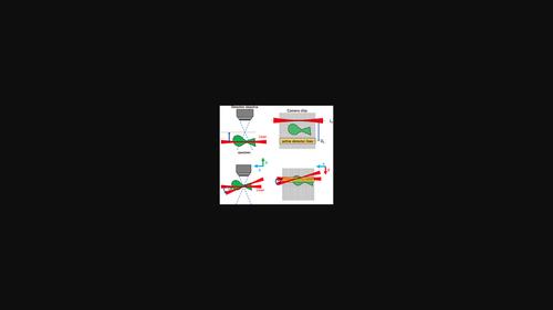
Light sheet fluorescence microscopy (LSFM) is an important tool in developmental biology. In this microscopy technique confocal line detection is often used to improve image contrast. To this end, the image of the illuminating scanned focused laser beam must be mapped onto a line detector. This is not trivial for long-term observations, since the spatial position of the laser beam and therefore its image on the detector may drift. The problem is aggravated in two-photon excitation LSFM, since pulsed laser light sources exhibit a lower laser beam pointing stability than continuous wave lasers. Here, we present a procedure for automatic synchronization between the excitation laser and detector, which does not require any additional hardware components and can therefore easily be integrated into existing systems. Since the recorded images are affected by noise, a specific, noise-tolerant focus metric was developed for calculating the relative displacement, which also allows for autofocusing in the detection direction. Furthermore, we developed an image analysis approach to determine a possible tilt of the excitation laser, which is executed in parallel to the autofocusing and enables the measurement of three solid angles. This allows to automatically correct for the tilting during a measurement. We demonstrated our approach by the observation of the migration of oligodendrocyte precursor cells in two-day-old fluorescent Tg(olig2:eGFP) reporter zebrafish larvae over a time span of more than 20 hours.
中文翻译:

使用共焦光片显微镜进行长期成像的自动检测器同步
光片荧光显微镜 (LSFM) 是发育生物学中的重要工具。在这种显微镜技术中,共焦线检测通常用于提高图像对比度。为此,照明扫描聚焦激光束的图像必须映射到线检测器上。这对于长期观察来说并不是微不足道的,因为激光束的空间位置及其在探测器上的图像可能会漂移。这个问题在双光子激发 LSFM 中更加严重,因为脉冲激光光源表现出比连续波激光器更低的激光束指向稳定性。在这里,我们提出了一个激发激光器和检测器之间自动同步的程序,它不需要任何额外的硬件组件,因此可以很容易地集成到现有系统中。由于记录的图像受到噪声的影响,因此开发了一种特定的、耐噪声的聚焦度量来计算相对位移,这也允许在检测方向上进行自动聚焦。此外,我们开发了一种图像分析方法来确定激发激光的可能倾斜度,该方法与自动聚焦并行执行并能够测量三个立体角。这允许在测量期间自动校正倾斜。我们通过观察两天大的荧光中少突胶质前体细胞的迁移来证明我们的方法 我们开发了一种图像分析方法来确定激发激光的可能倾斜度,该方法与自动聚焦并行执行并能够测量三个立体角。这允许在测量期间自动校正倾斜。我们通过观察两天大的荧光中少突胶质前体细胞的迁移来证明我们的方法 我们开发了一种图像分析方法来确定激发激光的可能倾斜度,该方法与自动聚焦并行执行并能够测量三个立体角。这允许在测量期间自动校正倾斜。我们通过观察两天大的荧光中少突胶质前体细胞的迁移来证明我们的方法Tg(olig2:eGFP)报告斑马鱼幼虫超过 20 小时的时间跨度。
更新日期:2022-08-20
中文翻译:
使用共焦光片显微镜进行长期成像的自动检测器同步
光片荧光显微镜 (LSFM) 是发育生物学中的重要工具。在这种显微镜技术中,共焦线检测通常用于提高图像对比度。为此,照明扫描聚焦激光束的图像必须映射到线检测器上。这对于长期观察来说并不是微不足道的,因为激光束的空间位置及其在探测器上的图像可能会漂移。这个问题在双光子激发 LSFM 中更加严重,因为脉冲激光光源表现出比连续波激光器更低的激光束指向稳定性。在这里,我们提出了一个激发激光器和检测器之间自动同步的程序,它不需要任何额外的硬件组件,因此可以很容易地集成到现有系统中。由于记录的图像受到噪声的影响,因此开发了一种特定的、耐噪声的聚焦度量来计算相对位移,这也允许在检测方向上进行自动聚焦。此外,我们开发了一种图像分析方法来确定激发激光的可能倾斜度,该方法与自动聚焦并行执行并能够测量三个立体角。这允许在测量期间自动校正倾斜。我们通过观察两天大的荧光中少突胶质前体细胞的迁移来证明我们的方法 我们开发了一种图像分析方法来确定激发激光的可能倾斜度,该方法与自动聚焦并行执行并能够测量三个立体角。这允许在测量期间自动校正倾斜。我们通过观察两天大的荧光中少突胶质前体细胞的迁移来证明我们的方法 我们开发了一种图像分析方法来确定激发激光的可能倾斜度,该方法与自动聚焦并行执行并能够测量三个立体角。这允许在测量期间自动校正倾斜。我们通过观察两天大的荧光中少突胶质前体细胞的迁移来证明我们的方法Tg(olig2:eGFP)报告斑马鱼幼虫超过 20 小时的时间跨度。


 京公网安备 11010802027423号