Diabetologia ( IF 8.2 ) Pub Date : 2022-08-12 , DOI: 10.1007/s00125-022-05779-2 Lars Krogvold 1, 2 , Angelo Genoni 3 , Anna Puggioni 3 , Daniela Campani 4 , Sarah J Richardson 5 , Christine S Flaxman 5 , Bjørn Edwin 6 , Trond Buanes 6 , Knut Dahl-Jørgensen 1, 7 , Antonio Toniolo 8
|
Aims/hypothesis
Enterovirus (EV) infection of pancreatic islet cells is one possible factor contributing to type 1 diabetes development. We have reported the presence of EV genome by PCR and of EV proteins by immunohistochemistry in pancreatic sections. Here we explore multiple human virus species in the Diabetes Virus Detection (DiViD) study cases using innovative methods, including virus passage in cell cultures.
Methods
Six recent-onset type 1 diabetes patients (age 24–35) were included in the DiViD study. Minimal pancreatic tail resection was performed under sterile conditions. Eleven live cases (age 43–83) of pancreatic carcinoma without diabetes served as control cases. In the present study, we used EV detection methods that combine virus growth in cell culture, gene amplification and detection of virus-coded proteins by immunofluorescence. Pancreas homogenates in cell culture medium were incubated with EV-susceptible cell lines for 3 days. Two to three blind passages were performed. DNA and RNA were extracted from both pancreas tissue and cell cultures. Real-time PCR was used for detecting 20 different viral agents other than EVs (six herpesviruses, human polyomavirus [BK virus and JC virus], parvovirus B19, hepatitis B virus, hepatitis C virus, hepatitis A virus, mumps, rubella, influenza A/B, parainfluenza 1–4, respiratory syncytial virus, astrovirus, norovirus, rotavirus). EV genomes were detected by endpoint PCR using five primer pairs targeting the partially conserved 5′ untranslated region genome region of the A, B, C and D species. Amplicons were sequenced. The expression of EV capsid proteins was evaluated in cultured cells using a panel of EV antibodies.
Results
Samples from six of six individuals with type 1 diabetes (cases) and two of 11 individuals without diabetes (control cases) contained EV genomes (p<0.05). In contrast, genomes of 20 human viruses other than EVs could be detected only once in an individual with diabetes (Epstein–Barr virus) and once in an individual without diabetes (parvovirus B19). EV detection was confirmed by immunofluorescence of cultured cells incubated with pancreatic extracts: viral antigens were expressed in the cytoplasm of approximately 1% of cells. Notably, infection could be transmitted from EV-positive cell cultures to uninfected cell cultures using supernatants filtered through 100 nm membranes, indicating that infectious agents of less than 100 nm were present in pancreases. Due to the slow progression of infection in EV-carrying cell cultures, cytopathic effects were not observed by standard microscopy but were recognised by measuring cell viability. Sequences of 5′ untranslated region amplicons were compatible with EVs of the B, A and C species. Compared with control cell cultures exposed to EV-negative pancreatic extracts, EV-carrying cell cultures produced significantly higher levels of IL-6, IL-8 and monocyte chemoattractant protein-1 (MCP1).
Conclusions/interpretation
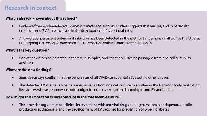
Sensitive assays confirm that the pancreases of all DiViD cases contain EVs but no other viruses. Analogous EV strains have been found in pancreases of two of 11 individuals without diabetes. The detected EV strains can be passaged in series from one cell culture to another in the form of poorly replicating live viruses encoding antigenic proteins recognised by multiple EV-specific antibodies. Thus, the early phase of type 1 diabetes is associated with a low-grade infection by EVs, but not by other viral agents.
Graphical abstract
中文翻译:
在 DiViD 研究中,在 1 型糖尿病发病时在人类胰腺中检测到活的肠道病毒,但没有检测到其他病毒
目标/假设
胰岛细胞的肠病毒 (EV) 感染是导致 1 型糖尿病发展的一个可能因素。我们已经通过 PCR 报告了 EV 基因组的存在,通过免疫组织化学报告了 EV 蛋白在胰腺切片中的存在。在这里,我们使用创新方法(包括细胞培养中的病毒传代)探索糖尿病病毒检测 (DiViD) 研究案例中的多种人类病毒。
方法
DiViD 研究包括六名新近发病的 1 型糖尿病患者(年龄 24-35 岁)。在无菌条件下进行最小胰尾切除术。11 例没有糖尿病的胰腺癌活体病例(43-83 岁)作为对照病例。在本研究中,我们使用的 EV 检测方法结合了细胞培养中的病毒生长、基因扩增和通过免疫荧光检测病毒编码蛋白。将细胞培养基中的胰腺匀浆与 EV 易感细胞系一起孵育 3 天。进行了两到三个盲传代。从胰腺组织和细胞培养物中提取 DNA 和 RNA。实时 PCR 用于检测 EV 以外的 20 种不同的病毒因子(六种疱疹病毒、人类多瘤病毒 [BK 病毒和 JC 病毒]、细小病毒 B19、乙型肝炎病毒、丙型肝炎病毒、甲型肝炎病毒、腮腺炎、风疹、A/B 型流感、副流感 1–4、呼吸道合胞病毒、星状病毒、诺瓦克病毒、轮状病毒)。使用针对 A、B、C 和 D 物种的部分保守的 5' 非翻译区基因组区域的五个引物对,通过终点 PCR 检测 EV 基因组。扩增子被测序。使用一组 EV 抗体在培养的细胞中评估 EV 衣壳蛋白的表达。
结果
来自 6 个 1 型糖尿病患者中的 6 个(病例)和 11 个没有糖尿病的个体中的 2 个(对照病例)的样本含有 EV 基因组(p<0.05)。相比之下,除 EV 外,20 种人类病毒的基因组只能在糖尿病患者(Epstein-Barr 病毒)和非糖尿病患者(细小病毒 B19)中检测到一次。通过用胰腺提取物孵育的培养细胞的免疫荧光证实了 EV 检测:病毒抗原在大约 1% 的细胞的细胞质中表达。值得注意的是,使用通过 100 nm 膜过滤的上清液,感染可以从 EV 阳性细胞培养物传播到未感染的细胞培养物,这表明胰腺中存在小于 100 nm 的感染因子。由于携带 EV 的细胞培养物中的感染进展缓慢,细胞病变效应无法通过标准显微镜观察到,但可以通过测量细胞活力来识别。5' 非翻译区扩增子的序列与 B、A 和 C 物种的 EV 兼容。与暴露于 EV 阴性胰腺提取物的对照细胞培养物相比,携带 EV 的细胞培养物产生显着更高水平的 IL-6、IL-8 和单核细胞趋化蛋白-1 (MCP1)。
结论/解释
敏感检测证实,所有 DiViD 病例的胰腺都含有 EV,但不含其他病毒。在 11 名没有糖尿病的人中有两人的胰腺中发现了类似的 EV 菌株。检测到的 EV 毒株可以从一种细胞培养物连续传代到另一种细胞培养物,其形式为复制能力较差的活病毒,其编码的抗原蛋白可被多种 EV 特异性抗体识别。因此,1 型糖尿病的早期阶段与 EV 的低度感染相关,但与其他病毒因子无关。


 京公网安备 11010802027423号