Journal of Clinical Monitoring and Computing ( IF 2.2 ) Pub Date : 2022-08-08 , DOI: 10.1007/s10877-022-00902-5 Andrea Costamagna 1, 2 , Irene Steinberg 2 , Emanuele Pivetta 3 , Pietro Arina 4 , Simona Veglia 5 , Luca Brazzi 1, 2 , Vito Fanelli 1, 2
|
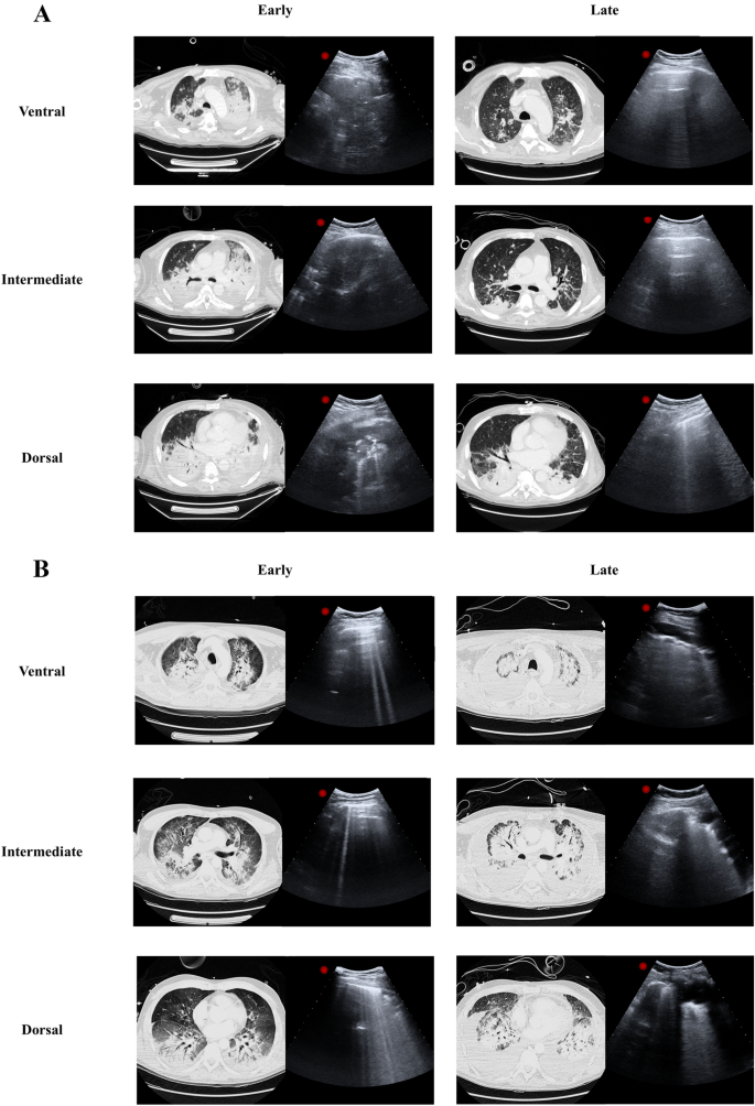
To evaluate whether lung ultrasound is reliable bedside tool to monitor changes of lung aeration at the early and late stages of ARDS. LUS was performed in ARDS patients that underwent at least two consecutive CT scan at ICU admission and at least 1 week after admission. Twelve fields were evaluated and graded from 0 (normal) to 3 (consolidation). Changes of LUS score in twelve fields (ΔLUStot) and in four ventral (ΔLUSV), intermediate (ΔLUSI) and dorsal (ΔLUSD) zones were calculated at each time points. Three categories were described: Improve (ΔLUS < 0), Equal (ΔLUS = 0) or Worse (ΔLUS > 0). LUS scores were correlated with total changes in lung CT aeration (ΔCTair) and with normally, poorly and not aerated regions (ΔCTnorm, ΔCTpoor and ΔCTnot, respectively). Eleven patients were enrolled. ΔLUStot had significant correlation with ΔCTair (r = − 0.74, p < 0.01). ΔLUSV, ΔLUSI and ΔLUSD showed significant correlations with ΔCTair (r = − 0.66, r = − 0.69, r = − 0.63, respectively; p < 0.05). Compared to Equal, Improve and Worse categories had significantly higher (p < 0.01) and lower (p < 0.05) ΔCTair values, respectively. Compared to Equal, Improve and Worse categories had lower (p < 0.01) and higher (p < 0.01) ΔCTnot values, respectively. LUS score had a good correlation with lung CT in detecting changes of lung aeration.
中文翻译:
肺部超声预测 ARDS 患者肺通气随时间变化的临床表现
评估肺部超声是否是监测 ARDS 早期和晚期肺通气变化的可靠床边工具。LUS 是在 ARDS 患者中进行的,这些患者在入住 ICU 时和入院后至少 1 周至少进行了两次连续 CT 扫描。对 12 个视野进行了评估,并从 0(正常)到 3(巩固)分级。在每个时间点计算十二个视野 (ΔLUS tot ) 和四个腹侧 (ΔLUS V )、中间 (ΔLUS I ) 和背侧 (ΔLUS D ) 区域的 LUS 评分变化。描述了三个类别:改善 (ΔLUS < 0)、相等 (ΔLUS = 0) 或更差 (ΔLUS > 0)。LUS 评分与肺 CT 通气的总变化相关(ΔCT空气) 以及正常、不良和未通气的区域(分别为 ΔCT norm、ΔCT poor和 ΔCT not)。登记了 11 名患者。ΔLUS tot与ΔCT空气显着相关(r = − 0.74,p < 0.01)。ΔLUS V、ΔLUS I和 ΔLUS D与 ΔCT空气显着相关(分别为 r = − 0.66、r = − 0.69、r = − 0.63;p < 0.05)。与“相等”、“改善”和“更差”类别相比,ΔCTair 值分别显着更高 (p < 0.01) 和更低 (p < 0.05)。与“相等”、“改善”和“更差”类别相比,ΔCT 较低 (p < 0.01) 和较高 (p < 0.01 )值,分别。LUS评分在检测肺通气变化方面与肺部CT有很好的相关性。


 京公网安备 11010802027423号