Diabetologia ( IF 8.2 ) Pub Date : 2022-07-25 , DOI: 10.1007/s00125-022-05756-9 Joseph J Castillo 1, 2 , Alfred C Aplin 1 , Daryl J Hackney 1 , Meghan F Hogan 1, 2 , Nathalie Esser 1, 2 , Andrew T Templin 1, 2 , Rehana Akter 1, 2 , Steven E Kahn 1, 2 , Daniel P Raleigh 3, 4 , Sakeneh Zraika 1, 2 , Rebecca L Hull 1, 2
|
Aims/hypothesis
The islet vasculature, including its constituent islet endothelial cells, is a key contributor to the microenvironment necessary for normal beta cell health and function. In type 2 diabetes, islet amyloid polypeptide (IAPP) aggregates, forming amyloid deposits that accumulate between beta cells and islet capillaries. This process is known to be toxic to beta cells but its impact on the islet vasculature has not previously been studied. Here, we report the first characterisation of the effects of IAPP aggregation on islet endothelial cells/capillaries using cell-based and animal models.
Methods
Primary and immortalised islet endothelial cells were treated with amyloidogenic human IAPP (hIAPP) alone or in the presence of the amyloid blocker Congo Red or the Toll-like receptor (TLR) 2/4 antagonist OxPAPc. Cell viability was determined0 along with mRNA and protein levels of inflammatory markers. Islet capillary abundance, morphology and pericyte coverage were determined in pancreases from transgenic mice with beta cell expression of hIAPP using conventional and confocal microscopy.
Results
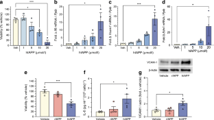
Aggregated hIAPP decreased endothelial cell viability in immortalised and primary islet endothelial cells (by 78% and 60%, respectively) and significantly increased expression of inflammatory markers Il6, Vcam1 and Edn1 mRNA relative to vehicle treatment in both cell types (p<0.05; n=4). Both cytotoxicity and the proinflammatory response were ameliorated by Congo Red (p<0.05; n=4); whereas TLR2/4-inhibition blocked inflammatory gene expression (p<0.05; n=6) without improving viability. Islets from high-fat-diet-fed amyloid-laden hIAPP transgenic mice also exhibited significantly increased expression of most markers of endothelial inflammation (p<0.05; n=5) along with decreased capillary density compared with non-transgenic littermates fed the same diet (p<0.01). Moreover, a 16% increase in capillary diameter was observed in amyloid-adjacent capillaries (p<0.01), accompanied by a doubling in pericyte structures positive for neuron-glial antigen 2 (p<0.001).
Conclusions/interpretation
Islet endothelial cells are susceptible to hIAPP-induced cytotoxicity and exhibit a TLR2/4-dependent proinflammatory response to aggregated hIAPP. Additionally, we observed amyloid-selective effects that decreased islet capillary density, accompanied by increased capillary diameter and increased pericyte number. Together, these data demonstrate that the islet vasculature is a target of the cytotoxic and proinflammatory effects of aggregated hIAPP that likely contribute to the detrimental effects of hIAPP aggregation on beta cell function and survival in type 2 diabetes.
Graphical abstract
中文翻译:
胰岛淀粉样蛋白多肽聚集对小鼠胰岛血管系统产生细胞毒性和促炎作用
目标/假设
胰岛血管系统,包括其组成的胰岛内皮细胞,是正常 β 细胞健康和功能所必需的微环境的关键贡献者。在 2 型糖尿病中,胰岛淀粉样多肽 (IAPP) 聚集,形成淀粉样沉积物,在 β 细胞和胰岛毛细血管之间积聚。已知该过程对 β 细胞有毒,但之前尚未研究其对胰岛脉管系统的影响。在这里,我们报告了使用基于细胞和动物模型的 IAPP 聚集对胰岛内皮细胞/毛细血管影响的首次表征。
方法
单独或在淀粉样蛋白阻断剂刚果红或 Toll 样受体 (TLR) 2/4 拮抗剂 OxPAPc 的存在下,原代和永生化胰岛内皮细胞用淀粉样蛋白生成人 IAPP (hIAPP) 进行处理。确定细胞活力以及炎症标志物的 mRNA 和蛋白质水平。使用常规和共聚焦显微镜在具有 hIAPP β 细胞表达的转基因小鼠的胰腺中测定胰岛毛细血管丰度、形态和周细胞覆盖率。
结果
聚集的 hIAPP 降低永生化和原代胰岛内皮细胞中的内皮细胞活力(分别降低 78% 和 60%),并且在两种细胞类型中相对于媒介物处理显着增加炎症标志物 Il6、Vcam1 和 Edn1 mRNA的表达( p < 0.05;n =4). 刚果红改善了细胞毒性和促炎反应(p <0.05;n = 4);而 TLR2/4 抑制可阻断炎症基因表达(p <0.05;n=6) 没有提高生存能力。与喂食相同饮食的非转基因同窝小鼠相比,高脂肪饮食喂养的富含淀粉样蛋白的 hIAPP 转基因小鼠的胰岛也表现出大多数内皮炎症标志物的表达显着增加(p <0.05;n = 5)以及毛细血管密度降低(p <0.01)。此外,在与淀粉样蛋白相邻的毛细血管中观察到毛细血管直径增加了 16% ( p <0.01),同时神经元-神经胶质抗原 2 阳性的周细胞结构加倍 ( p <0.001)。
结论/解释
胰岛内皮细胞对 hIAPP 诱导的细胞毒性敏感,并对聚集的 hIAPP 表现出 TLR2/4 依赖性促炎反应。此外,我们观察到淀粉样蛋白选择性作用会降低胰岛毛细血管密度,并伴有毛细血管直径增加和周细胞数量增加。总之,这些数据表明胰岛脉管系统是聚集的 hIAPP 的细胞毒性和促炎作用的靶点,这可能导致 hIAPP 聚集对 2 型糖尿病 β 细胞功能和存活的不利影响。


 京公网安备 11010802027423号