Journal of Molecular Histology ( IF 3.2 ) Pub Date : 2022-07-21 , DOI: 10.1007/s10735-022-10084-8 Huanhuan Li 1 , Xueqiang Wu 2 , Dingfang Bu 3 , Lihua Wang 2 , Xueju Xu 1 , Yingchao Wang 1 , Yufeng Liu 1 , Ping Zhu 3
|
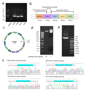
High Mobility Group Chromosomal Protein N2 (HMGN2) can recognize tumor cells and enhance the anti-tumor effect of immune cells. This study aimed to establish a lentiviral vector of recombinant HMGN2 gene, establish recombinant T cells (HMGN2-T cells), and observe their anti-tumor effects. Total RNA was isolated from peripheral blood mononuclear cells. HMGN2, cluster of differentiation (CD) 8 A, CD28, CD137, and CD3ζ genes were amplified and connected. Jurkat cells were transfected with the recombinant lentivirus vector. The viability, apoptosis, and cell cycle of HMGN2-T cells were detected using Cell Counting Kit-8 assay and flow cytometry. The co-culture was performed by adding HMGN2-T cells to tumor cells with different effect-to-target (E:T) ratios. The cytotoxic activity was measured by lactate dehydrogenase (LDH) releasing assay. The sequences of HMGN2, CD8A, CD28, CD137, and CD3ζ gene plasmids were confirmed using gene sequencing. After the lentiviral transfection for 72 h, green fluorescence cells (HMGN2-T cells) could be seen. Cell viability and apoptosis were increased in HMGN2-T cells. The cytokine levels of interleukin 2 (IL-2) and tumor necrosis factor α (TNF-α) increased in cell supernatants of HMGN2-T cells. The percentage of G0/G1 phase cells was lower, the rate of S phase cells was higher in HMGN2-T cells than control cells. The co-culture of HMGN2-T cells and tumor cells could promote the cytokines’ release. The LDH level was increased with the elevation of E:T ratios. In conclusion, the HMGN2-T cells were well-established and have the effect of secreting cytokines and killing tumor cells.
中文翻译:
重组 jurkat 细胞(HMGN2-T 细胞)分泌细胞因子并抑制肿瘤细胞的生长
高迁移率群染色体蛋白N2(HMGN2)可以识别肿瘤细胞,增强免疫细胞的抗肿瘤作用。本研究旨在建立重组HMGN2基因的慢病毒载体,建立重组T细胞(HMGN2-T细胞),并观察其抗肿瘤作用。从外周血单核细胞中分离总RNA。HMGN2、分化簇 (CD) 8 A、CD28、CD137 和 CD3ζ 基因被扩增和连接。用重组慢病毒载体转染 Jurkat 细胞。使用 Cell Counting Kit-8 检测和流式细胞术检测 HMGN2-T 细胞的活力、凋亡和细胞周期。通过将 HMGN2-T 细胞添加到具有不同效果与靶标 (E:T) 比率的肿瘤细胞中来进行共培养。通过乳酸脱氢酶(LDH)释放测定法测量细胞毒活性。HMGN2、CD8A、CD28、CD137和CD3ζ基因质粒的序列通过基因测序得到证实。慢病毒转染72 h后,可见绿色荧光细胞(HMGN2-T细胞)。HMGN2-T 细胞中的细胞活力和凋亡增加。HMGN2-T 细胞上清液中白细胞介素 2 (IL-2) 和肿瘤坏死因子 α (TNF-α) 的细胞因子水平升高。HMGN2-T细胞中G0/G1期细胞百分比较低,S期细胞率高于对照细胞。HMGN2-T细胞与肿瘤细胞共培养可促进细胞因子的释放。LDH 水平随着 E:T 比值的升高而增加。综上所述,HMGN2-T细胞建立良好,具有分泌细胞因子和杀伤肿瘤细胞的作用。和 CD3ζ 基因质粒通过基因测序得到证实。慢病毒转染72 h后,可见绿色荧光细胞(HMGN2-T细胞)。HMGN2-T 细胞中的细胞活力和凋亡增加。HMGN2-T 细胞上清液中白细胞介素 2 (IL-2) 和肿瘤坏死因子 α (TNF-α) 的细胞因子水平升高。HMGN2-T细胞中G0/G1期细胞百分比较低,S期细胞率高于对照细胞。HMGN2-T细胞与肿瘤细胞共培养可促进细胞因子的释放。LDH 水平随着 E:T 比值的升高而增加。综上所述,HMGN2-T细胞建立良好,具有分泌细胞因子和杀伤肿瘤细胞的作用。和 CD3ζ 基因质粒通过基因测序得到证实。慢病毒转染72 h后,可见绿色荧光细胞(HMGN2-T细胞)。HMGN2-T 细胞中的细胞活力和凋亡增加。HMGN2-T 细胞上清液中白细胞介素 2 (IL-2) 和肿瘤坏死因子 α (TNF-α) 的细胞因子水平升高。HMGN2-T细胞中G0/G1期细胞百分比较低,S期细胞率高于对照细胞。HMGN2-T细胞与肿瘤细胞共培养可促进细胞因子的释放。LDH 水平随着 E:T 比值的升高而增加。综上所述,HMGN2-T细胞建立良好,具有分泌细胞因子和杀伤肿瘤细胞的作用。可见绿色荧光细胞(HMGN2-T 细胞)。HMGN2-T 细胞中的细胞活力和凋亡增加。HMGN2-T 细胞上清液中白细胞介素 2 (IL-2) 和肿瘤坏死因子 α (TNF-α) 的细胞因子水平升高。HMGN2-T细胞中G0/G1期细胞百分比较低,S期细胞率高于对照细胞。HMGN2-T细胞与肿瘤细胞共培养可促进细胞因子的释放。LDH 水平随着 E:T 比值的升高而增加。综上所述,HMGN2-T细胞建立良好,具有分泌细胞因子和杀伤肿瘤细胞的作用。可见绿色荧光细胞(HMGN2-T 细胞)。HMGN2-T 细胞中的细胞活力和凋亡增加。HMGN2-T 细胞上清液中白细胞介素 2 (IL-2) 和肿瘤坏死因子 α (TNF-α) 的细胞因子水平升高。HMGN2-T细胞中G0/G1期细胞百分比较低,S期细胞率高于对照细胞。HMGN2-T细胞与肿瘤细胞共培养可促进细胞因子的释放。LDH 水平随着 E:T 比值的升高而增加。综上所述,HMGN2-T细胞建立良好,具有分泌细胞因子和杀伤肿瘤细胞的作用。HMGN2-T细胞中G0/G1期细胞百分比较低,S期细胞率高于对照细胞。HMGN2-T细胞与肿瘤细胞共培养可促进细胞因子的释放。LDH 水平随着 E:T 比值的升高而增加。综上所述,HMGN2-T细胞建立良好,具有分泌细胞因子和杀伤肿瘤细胞的作用。HMGN2-T细胞中G0/G1期细胞百分比较低,S期细胞率高于对照细胞。HMGN2-T细胞与肿瘤细胞共培养可促进细胞因子的释放。LDH 水平随着 E:T 比值的升高而增加。综上所述,HMGN2-T细胞建立良好,具有分泌细胞因子和杀伤肿瘤细胞的作用。


 京公网安备 11010802027423号