Cell Metabolism ( IF 29.0 ) Pub Date : 2022-07-19 , DOI: 10.1016/j.cmet.2022.06.012 Pieter Goossens 1 , Chang Lu 1 , Jianhua Cao 2 , Marion J Gijbels 3 , Joël M H Karel 4 , Erwin Wijnands 5 , Britt S R Claes 2 , Gregorio E Fazzi 1 , Tim F E Hendriks 2 , Kristiaan Wouters 6 , Evgueni Smirnov 4 , Marc J M van Zandvoort 7 , Benjamin Balluff 2 , Eva Cuypers 2 , Marjo M P C Donners 1 , Ron M A Heeren 2 , Erik A L Biessen 8
|
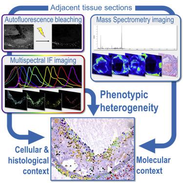
Cells often adopt different phenotypes, dictated by tissue-specific or local signals such as cell-cell and cell-matrix contacts or molecular micro-environment. This holds in extremis for macrophages with their high phenotypic plasticity. Their broad range of functions, some even opposing, reflects their heterogeneity, and a multitude of subsets has been described in different tissues and diseases. Such micro-environmental imprint cannot be adequately studied by single-cell applications, as cells are detached from their context, while histology-based assessment lacks the phenotypic depth due to limitations in marker combination. Here, we present a novel, integrative approach in which 15-color multispectral imaging allows comprehensive cell classification based on multi-marker expression patterns, followed by downstream analysis pipelines to link their phenotypes to contextual, micro-environmental cues, such as their cellular (“community”) and metabolic (“local lipidome”) niches in complex tissue. The power of this approach is illustrated for myeloid subsets and associated lipid signatures in murine atherosclerotic plaque.
中文翻译:
整合多重免疫荧光和质谱成像以绘制代谢和细胞环境中的骨髓异质性
细胞通常采用不同的表型,由组织特异性或局部信号决定,例如细胞-细胞和细胞-基质接触或分子微环境。这在极端情况下对于具有高表型可塑性的巨噬细胞。它们广泛的功能,有些甚至是相反的,反映了它们的异质性,并且在不同的组织和疾病中描述了许多子集。这种微环境印记无法通过单细胞应用进行充分研究,因为细胞与其环境分离,而基于组织学的评估由于标记组合的限制而缺乏表型深度。在这里,我们提出了一种新颖的综合方法,其中 15 色多光谱成像允许基于多标记表达模式进行全面的细胞分类,然后通过下游分析管道将它们的表型与上下文、微环境线索(例如它们的细胞)联系起来。复杂组织中的“社区”)和代谢(“局部脂质组”)生态位。


 京公网安备 11010802027423号