Diabetologia ( IF 8.2 ) Pub Date : 2022-07-19 , DOI: 10.1007/s00125-022-05719-0 Emma M Lessieur 1 , Haitao Liu 2, 3 , Aicha Saadane 1 , Yunpeng Du 1 , Jianying Kiser 1 , Timothy S Kern 1, 4
|
Aims/hypothesis
Induction of intercellular adhesion molecule-1 (ICAM-1) has been implicated in the development of macrovascular and microvascular diseases such as diabetic retinopathy. Lesions of diabetic retinopathy are unique to the retina but the reason for this is unclear, as all tissues are exposed to the same hyperglycaemic insult. We tested whether diabetes induces ICAM-1 on the luminal surface of endothelial cells to a greater extent in the retina than in other tissues and the role of vision itself in that induction.
Methods
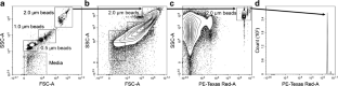
Experimental diabetes was induced in C57Bl/6J, P23H opsin mutant and Gnat1−/− × Gnat2−/− double knockout mice using streptozotocin. The relative abundance of ICAM-1 on the luminal surface of endothelial cells in retina and other tissues was determined by conjugating anti-ICAM-1 antibodies to fluorescent microspheres (2 μm), injecting them intravenously and allowing them to circulate for 30 min. After transcardial perfusion, quantification of microspheres adherent to the endothelium in tissues throughout the body was carried out by fluorescent microscopy or flow cytometry. Mice injected with lipopolysaccharide (LPS) were used as positive controls. The difference in leucostasis between retinal and non-retinal vasculature was evaluated.
Results
Diabetes significantly increased ICAM-1-mediated adherence of microspheres to retinal microvessels by almost threefold, independent of sex. In contrast, diabetes had a much smaller effect on endothelial ICAM-1 in other tissues, and more tissues showed a significant induction of endothelial ICAM-1 with LPS than with diabetes. The diabetes-induced increase in endothelial ICAM-1 in retinal vasculature was inhibited by blocking phototransduction in photoreceptor cells. Diabetes significantly increased leucostasis in the retina by threefold compared with a non-ocular tissue (cremaster).
Conclusions/interpretation
The diabetes-induced upregulation of ICAM-1 on the luminal surface of the vascular endothelium varies considerably among tissues and is highest in the retina. Induction of ICAM-1 on retinal vascular endothelial cells in diabetes is influenced by vision-related processes in photoreceptor cells. The unique presence of photoreceptors in the retina might contribute to the greater susceptibility of this tissue to vascular disease in diabetes.
Graphical abstract
中文翻译:
糖尿病患者视网膜内皮细胞管腔表面的 ICAM-1 诱导程度高于其他组织
目标/假设
细胞间粘附分子 1 (ICAM-1) 的诱导与糖尿病视网膜病变等大血管和微血管疾病的发生有关。糖尿病视网膜病变的病变是视网膜所特有的,但其原因尚不清楚,因为所有组织都暴露于相同的高血糖损伤。我们测试了糖尿病是否会比其他组织更大程度地在视网膜内皮细胞管腔表面诱导 ICAM-1,以及视觉本身在该诱导中的作用。
方法
使用链脲佐菌素在 C57Bl/6J、 P23H视蛋白突变体和Gnat1 −/− × Gnat2 −/−双敲除小鼠中诱导实验性糖尿病。通过将抗 ICAM-1 抗体与荧光微球 (2 μm) 结合,静脉注射并循环 30 分钟,测定视网膜和其他组织内皮细胞管腔表面上 ICAM-1 的相对丰度。经心灌注后,通过荧光显微镜或流式细胞术对全身组织中粘附于内皮的微球进行定量。注射脂多糖(LPS)的小鼠用作阳性对照。评估视网膜和非视网膜脉管系统之间白细胞淤积的差异。
结果
糖尿病使 ICAM-1 介导的微球与视网膜微血管的粘附显着增加了近三倍,与性别无关。相比之下,糖尿病对其他组织中的内皮 ICAM-1 的影响要小得多,并且与糖尿病相比,更多的组织显示 LPS 对内皮 ICAM-1 的显着诱导。通过阻断感光细胞中的光转导,可以抑制糖尿病引起的视网膜血管中内皮 ICAM-1 的增加。与非眼部组织(提睾肌)相比,糖尿病使视网膜中的白细胞淤滞显着增加了三倍。
结论/解释
糖尿病引起的血管内皮管腔表面 ICAM-1 的上调在不同组织中差异很大,并且在视网膜中最高。糖尿病患者视网膜血管内皮细胞上 ICAM-1 的诱导受到感光细胞中视觉相关过程的影响。视网膜中光感受器的独特存在可能导致该组织更容易患糖尿病血管疾病。


 京公网安备 11010802027423号