Nature Biomedical Engineering ( IF 28.1 ) Pub Date : 2022-07-14 , DOI: 10.1038/s41551-022-00906-1 Ruiqing Ni 1, 2, 3 , Zhenyue Chen 1, 4 , Xosé Luís Deán-Ben 1, 4 , Fabian F Voigt 2, 5 , Daniel Kirschenbaum 6 , Gloria Shi 1 , Alessia Villois 7 , Quanyu Zhou 1, 4 , Alessandro Crimi 6 , Paolo Arosio 7 , Roger M Nitsch 2, 3 , K Peter R Nilsson 8 , Adriano Aguzzi 2, 6 , Fritjof Helmchen 2, 5 , Jan Klohs 1, 2 , Daniel Razansky 1, 2, 4
|
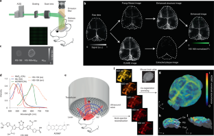
Deposits of amyloid-β (Aβ) in the brains of rodents can be analysed by invasive intravital microscopy on a submillimetre scale, or via whole-brain images from modalities lacking the resolution or molecular specificity to accurately characterize Aβ pathologies. Here we show that large-field multifocal illumination fluorescence microscopy and panoramic volumetric multispectral optoacoustic tomography can be combined to longitudinally assess Aβ deposits in transgenic mouse models of Alzheimer’s disease. We used fluorescent Aβ-targeted probes (the luminescent conjugated oligothiophene HS-169 and the oxazine-derivative AOI987) to transcranially detect Aβ deposits in the cortex of APP/PS1 and arcAβ mice with single-plaque resolution (8 μm) and across the whole brain (including the hippocampus and the thalamus, which are inaccessible by conventional intravital microscopy) at sub-150 μm resolutions. Two-photon microscopy, light-sheet microscopy and immunohistochemistry of brain-tissue sections confirmed the specificity and regional distributions of the deposits. High-resolution multiscale optical and optoacoustic imaging of Aβ deposits across the entire brain in rodents thus facilitates the in vivo study of Aβ accumulation by brain region and by animal age and strain.
中文翻译:
小鼠β淀粉样蛋白沉积物的多尺度光学和光声成像
啮齿动物大脑中淀粉样蛋白-β (Aβ) 的沉积物可以通过亚毫米级的侵入性活体显微镜进行分析,或者通过来自缺乏分辨率或分子特异性的方式的全脑图像来准确表征 Aβ 病理。在这里,我们展示了大视野多焦点照明荧光显微镜和全景体积多光谱光声断层扫描可以结合起来纵向评估阿尔茨海默病转基因小鼠模型中的 Aβ 沉积物。我们使用荧光 Aβ 靶向探针(发光共轭低聚噻吩 HS-169 和恶嗪衍生物 AOI987)经颅检测 APP/PS1 和 arcAβ 小鼠皮质中的 Aβ 沉积物,具有单斑块分辨率(8 μm)和整个大脑(包括海马体和丘脑,在亚 150 μm 的分辨率下,传统的活体显微镜无法访问。脑组织切片的双光子显微镜、光片显微镜和免疫组织化学证实了沉积物的特异性和区域分布。因此,啮齿动物整个大脑中 Aβ 沉积物的高分辨率多尺度光学和光声成像有助于在体内研究按大脑区域和动物年龄和应变的 Aβ 积累。


 京公网安备 11010802027423号