European Journal of Nuclear Medicine and Molecular Imaging ( IF 9.1 ) Pub Date : 2022-07-13 , DOI: 10.1007/s00259-022-05902-w Joey Roosen 1 , Lovisa E L Westlund Gotby 1 , Mark J Arntz 1 , Jurgen J Fütterer 1 , Marcel J R Janssen 1 , Mark W Konijnenberg 1, 2 , Meike W M van Wijk 1 , Christiaan G Overduin 1 , J Frank W Nijsen 1
|
Purpose
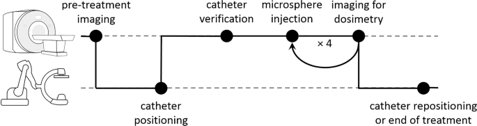
Transarterial radioembolization (TARE) is a treatment for liver tumours based on injection of radioactive microspheres in the hepatic arterial system. It is crucial to achieve a maximum tumour dose for an optimal treatment response, while minimizing healthy liver dose to prevent toxicity. There is, however, no intraprocedural feedback on the dose distribution, as nuclear imaging can only be performed after treatment. As holmium-166 (166Ho) microspheres can be quantified with MRI, we investigate the feasibility and safety of performing 166Ho TARE within an MRI scanner and explore the potential of intraprocedural MRI-based dosimetry.
Methods
Six patients were treated with 166Ho TARE in a hybrid operating room. Per injection position, a microcatheter was placed under angiography guidance, after which patients were transported to an adjacent 3-T MRI system. After MRI confirmation of unchanged catheter location, 166Ho microspheres were injected in four fractions, consisting of 10%, 30%, 30% and 30% of the planned activity, alternated with holmium-sensitive MRI acquisition to assess the microsphere distribution. After the procedures, MRI-based dose maps were calculated from each intraprocedural image series using a dedicated dosimetry software package for 166Ho TARE.
Results
Administration of 166Ho microspheres within the MRI scanner was feasible in 9/11 (82%) injection positions. Intraprocedural holmium-sensitive MRI allowed for tumour dosimetry in 18/19 (95%) of treated tumours. Two CTCAE grade 3–4 toxicities were observed, and no adverse events were attributed to treatment in the MRI. Towards the last fraction, 4/18 tumours exhibited signs of saturation, while in 14/18 tumours, the microsphere uptake patterns did not deviate from the linear trend.
Conclusion
This study demonstrated feasibility and preliminary safety of a first in-human application of TARE within a clinical MRI system. Intraprocedural MRI-based dosimetry enabled dynamic insight in the microsphere distribution during TARE. This proof of concept yields unique possibilities to better understand microsphere distribution in vivo and to potentially optimize treatment efficacy through treatment personalization.
Registration
Clinicaltrials.gov, identifier NCT04269499, registered on February 13, 2020 (retrospectively registered).
中文翻译:
用钬 166 微球 (EMERITUS-1) 对肝肿瘤进行经动脉放射栓塞术期间基于 MRI 的程序内剂量测定:针对适应性图像控制治疗实施的 I 期试验
目的
经动脉放射栓塞术 (TARE) 是一种基于在肝动脉系统中注射放射性微球体的肝肿瘤治疗方法。实现最佳治疗反应的最大肿瘤剂量至关重要,同时尽量减少健康肝脏剂量以防止毒性。然而,没有关于剂量分布的程序内反馈,因为核成像只能在治疗后进行。由于钬 166 ( 166 Ho) 微球可以用 MRI 进行量化,我们调查了在 MRI 扫描仪内执行166 Ho TARE 的可行性和安全性,并探索了基于 MRI 的程序内剂量测定的潜力。
方法
六名患者在混合手术室接受了166 Ho TARE 治疗。在每个注射位置,在血管造影引导下放置一根微导管,然后将患者运送到相邻的 3-T MRI 系统。在 MRI 确认导管位置不变后,将166 个Ho 微球分为四个部分注射,分别为计划活动的 10%、30%、30% 和 30%,交替进行钬敏感 MRI 采集以评估微球分布。手术后,使用用于166 Ho TARE的专用剂量学软件包从每个手术过程中的图像系列计算基于 MRI 的剂量图。
结果
在 MRI 扫描仪内管理166 Ho 微球在 9/11 (82%) 注射位置是可行的。术中钬敏感性 MRI 允许对 18/19 (95%) 的治疗肿瘤进行肿瘤剂量测定。观察到两个 CTCAE 3-4 级毒性,并且没有不良事件归因于 MRI 中的治疗。在最后一部分,4/18 的肿瘤表现出饱和迹象,而在 14/18 的肿瘤中,微球摄取模式没有偏离线性趋势。
结论
这项研究证明了在临床 MRI 系统中首次在人体中应用 TARE 的可行性和初步安全性。基于 MRI 的程序内剂量测定能够动态洞察 TARE 期间的微球分布。这种概念验证产生了独特的可能性,可以更好地了解微球在体内的分布,并可能通过个性化治疗来优化治疗效果。
登记
Clinicaltrials.gov,标识符 NCT04269499,于 2020 年 2 月 13 日注册(追溯注册)。


 京公网安备 11010802027423号