Medical Image Analysis ( IF 10.9 ) Pub Date : 2022-07-09 , DOI: 10.1016/j.media.2022.102528 Xiahai Zhuang 1 , Jiahang Xu 1 , Xinzhe Luo 1 , Chen Chen 2 , Cheng Ouyang 2 , Daniel Rueckert 2 , Victor M Campello 3 , Karim Lekadir 3 , Sulaiman Vesal 4 , Nishant RaviKumar 4 , Yashu Liu 5 , Gongning Luo 5 , Jingkun Chen 6 , Hongwei Li 7 , Buntheng Ly 8 , Maxime Sermesant 8 , Holger Roth 9 , Wentao Zhu 9 , Jiexiang Wang 10 , Xinghao Ding 10 , Xinyue Wang 11 , Sen Yang 12 , Lei Li 13
|
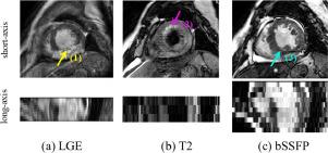
Accurate computing, analysis and modeling of the ventricles and myocardium from medical images are important, especially in the diagnosis and treatment management for patients suffering from myocardial infarction (MI). Late gadolinium enhancement (LGE) cardiac magnetic resonance (CMR) provides an important protocol to visualize MI. However, compared with the other sequences LGE CMR images with gold standard labels are particularly limited. This paper presents the selective results from the Multi-Sequence Cardiac MR (MS-CMR) Segmentation challenge, in conjunction with MICCAI 2019. The challenge offered a data set of paired MS-CMR images, including auxiliary CMR sequences as well as LGE CMR, from 45 patients who underwent cardiomyopathy. It was aimed to develop new algorithms, as well as benchmark existing ones for LGE CMR segmentation focusing on myocardial wall of the left ventricle and blood cavity of the two ventricles. In addition, the paired MS-CMR images could enable algorithms to combine the complementary information from the other sequences for the ventricle segmentation of LGE CMR. Nine representative works were selected for evaluation and comparisons, among which three methods are unsupervised domain adaptation (UDA) methods and the other six are supervised. The results showed that the average performance of the nine methods was comparable to the inter-observer variations. Particularly, the top-ranking algorithms from both the supervised and UDA methods could generate reliable and robust segmentation results. The success of these methods was mainly attributed to the inclusion of the auxiliary sequences from the MS-CMR images, which provide important label information for the training of deep neural networks. The challenge continues as an ongoing resource, and the gold standard segmentation as well as the MS-CMR images of both the training and test data are available upon registration via its homepage (www.sdspeople.fudan.edu.cn/zhuangxiahai/0/mscmrseg/).
中文翻译:
晚期钆增强 MRI 的心脏分割:来自多序列心脏 MR 分割挑战的基准研究
对医学图像中的心室和心肌进行精确计算、分析和建模非常重要,尤其是在心肌梗死(MI)患者的诊断和治疗管理中。晚期钆增强 (LGE) 心脏磁共振 (CMR) 提供了可视化 MI 的重要协议。然而,与其他序列相比,带有金标准标签的 LGE CMR 图像特别有限。本文结合 MICCAI 2019 介绍了多序列心脏 MR (MS-CMR) 分割挑战的选择性结果。该挑战提供了成对的 MS-CMR 图像数据集,包括辅助 CMR 序列以及 LGE CMR,来自 45 名患有心肌病的患者。它的目的是开发新的算法,以及以左心室心肌壁和两心室血腔为重点的 LGE CMR 分割的现有基准。此外,配对的 MS-CMR 图像可以使算法结合来自其他序列的互补信息,用于 LGE CMR 的心室分割。选取了 9 个具有代表性的作品进行评估和比较,其中 3 个方法是无监督领域自适应(UDA)方法,另外 6 个是有监督的。结果表明,九种方法的平均性能与观察者间的差异相当。特别是,来自监督和 UDA 方法的排名靠前的算法可以生成可靠且稳健的分割结果。这些方法的成功主要归功于包含来自 MS-CMR 图像的辅助序列,这为深度神经网络的训练提供了重要的标签信息。挑战作为一个持续的资源继续进行,黄金标准分割以及训练和测试数据的 MS-CMR 图像可在其主页(www.sdspeople.fudan.edu.cn/zhuangxiahai/0/)注册后获得mscmrseg/).


 京公网安备 11010802027423号