European Journal of Nuclear Medicine and Molecular Imaging ( IF 9.1 ) Pub Date : 2022-07-05 , DOI: 10.1007/s00259-022-05868-9 Gregory T Kennedy 1 , Feredun S Azari 1 , Elizabeth Bernstein 1 , Bilal Nadeem 1 , Ashley Chang 1 , Alix Segil 1 , Neil Sullivan 1 , Emmanuel Encarnado 1 , Charuhas Desphande 2 , John C Kucharczuk 1 , Kaela Leonard 3 , Philip S Low 4 , Silvia Chen 5 , Aline Criton 3 , Sunil Singhal 1
|
Background
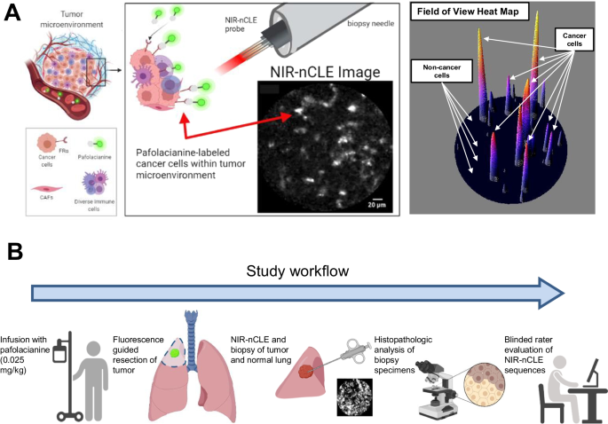
The diagnostic yield of biopsies of solitary pulmonary nodules (SPNs) is low, particularly in sub-solid lesions. We developed a method (NIR-nCLE) to achieve cellular level cancer detection during biopsy by integrating (i) near-infrared (NIR) imaging using a cancer-targeted tracer (pafolacianine), and (ii) a flexible NIR confocal laser endomicroscopy (CLE) system that can fit within a biopsy needle. Our goal was to assess the diagnostic accuracy of NIR-nCLE ex vivo in SPNs.
Methods
Twenty patients with SPNs were preoperatively infused with pafolacianine. Following resection, specimens were inspected to identify the lesion of interest. NIR-nCLE imaging followed by tissue biopsy was performed within the lesion and in normal lung tissue. All imaging sequences (n = 115) were scored by 5 blinded raters on the presence of fluorescent cancer cells and compared to diagnoses by a thoracic pathologist.
Results
Most lesions (n = 15, 71%) were adenocarcinoma-spectrum malignancies, including 7 ground glass opacities (33%). Mean fluorescence intensity (MFI) by NIR-nCLE for tumor biopsy was 20.6 arbitrary units (A.U.) and mean MFI for normal lung was 6.4 A.U. (p < 0.001). Receiver operating characteristic analysis yielded a high area under the curve for MFI (AUC = 0.951). Blinded raters scored the NIR-nCLE sequences on the presence of fluorescent cancer cells with sensitivity and specificity of 98% and 97%, respectively. Overall diagnostic accuracy was 97%. The inter-observer agreement of the five raters was excellent (κ = 0.95).
Conclusions
NIR-nCLE allows sensitive and specific detection of cancer cells in SPNs. This technology has far-reaching implications for diagnostic needle biopsies and intraprocedural decision-making.
中文翻译:
在活检过程中靶向检测癌细胞可以实时诊断肺结节
背景
孤立性肺结节 (SPN) 活检的诊断率很低,尤其是亚实性病变。我们开发了一种方法 (NIR-nCLE),通过整合 (i) 使用癌症靶向示踪剂 (pafolacianine) 的近红外 (NIR) 成像和 (ii) 灵活的 NIR 共聚焦激光显微术,在活检期间实现细胞水平的癌症检测。 CLE) 系统,可以安装在活检针内。我们的目标是评估 NIR-nCLE 离体在 SPN 中的诊断准确性。
方法
20 名 SPN 患者术前输注了 pafolacianine。切除后,检查标本以确定感兴趣的病变。在病变和正常肺组织中进行 NIR-nCLE 成像,然后进行组织活检。所有成像序列 ( n = 115) 由 5 名盲法评分者根据荧光癌细胞的存在进行评分,并与胸腔病理学家的诊断进行比较。
结果
大多数病变 ( n = 15, 71%) 是腺癌谱系恶性肿瘤,包括 7 个磨玻璃影 (33%)。NIR-nCLE 的肿瘤活检平均荧光强度 (MFI) 为 20.6 任意单位 (AU),正常肺的平均 MFI 为 6.4 AU ( p < 0.001)。接受者操作特征分析产生了 MFI 曲线下的高面积(AUC = 0.951)。盲法评分者根据荧光癌细胞的存在对 NIR-nCLE 序列进行评分,灵敏度和特异性分别为 98% 和 97%。总体诊断准确率为 97%。五名评估者的观察者间一致性非常好 ( κ = 0.95)。
结论
NIR-nCLE 允许对 SPN 中的癌细胞进行灵敏和特异性检测。该技术对诊断性穿刺活检和术中决策具有深远影响。


 京公网安备 11010802027423号