当前位置:
X-MOL 学术
›
Anal. Chem.
›
论文详情
Our official English website, www.x-mol.net, welcomes your feedback! (Note: you will need to create a separate account there.)
Neurotransmitter Readily Escapes Detection at the Opposing Microelectrode Surface in Typical Amperometric Measurements of Exocytosis at Single Cells
Analytical Chemistry ( IF 7.4 ) Pub Date : 2022-06-24 , DOI: 10.1021/acs.analchem.2c00060 Gregory S McCarty , Lars E Dunaway , J Dylan Denison , Leslie A Sombers
Analytical Chemistry ( IF 7.4 ) Pub Date : 2022-06-24 , DOI: 10.1021/acs.analchem.2c00060 Gregory S McCarty , Lars E Dunaway , J Dylan Denison , Leslie A Sombers
|
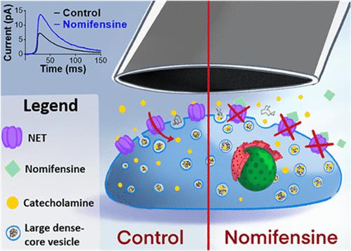
For decades, carbon-fiber microelectrodes have been used in amperometric measurements of neurotransmitter release at a wide variety of cell types, providing a tremendous amount of valuable information on the mechanisms involved in dense-core vesicle fusion. The electroactive molecules that are released can be detected at the opposing microelectrode surface, allowing for precise quantification as well as detailed kinetic information on the stages of neurotransmitter release. However, it remains unclear how much of the catecholamine that is released into the artificial synapse escapes detection. This work examines two separate mechanisms by which released neurotransmitter goes undetected in a typical amperometric measurement. First, diffusional loss is assessed by monitoring exocytosis at single bovine chromaffin cells using carbon-fiber microelectrodes fabricated in a recessed (cavity) geometry. This creates a microsampling vial that minimizes diffusional loss of analyte prior to detection. More molecules were detected per exocytotic release event when using a recessed cavity sensor as compared to the conventional configuration. In addition, pharmacological inhibition of the norepinephrine transporter (NET), which serves to remove catecholamine from the extracellular space, increased both the size and the time course of individual amperometric events. Overall, this study characterizes distinct physical and biological mechanisms by which released neurotransmitter escapes detection at the opposing microelectrode surface, while also revealing an important role for the NET in “presynaptic” modulation of neurotransmitter release.
中文翻译:

在单细胞胞吐作用的典型安培测量中,神经递质很容易逃脱相对微电极表面的检测
几十年来,碳纤维微电极已用于对多种细胞类型的神经递质释放进行电流测量,为致密核囊泡融合机制提供了大量有价值的信息。释放的电活性分子可以在相对的微电极表面进行检测,从而可以进行精确的定量以及神经递质释放阶段的详细动力学信息。然而,目前尚不清楚释放到人工突触中的儿茶酚胺有多少逃脱了检测。这项工作研究了两种不同的机制,通过这两种机制,释放的神经递质在典型的电流测量中未被检测到。首先,通过使用凹进(腔)几何形状制造的碳纤维微电极监测单个牛嗜铬细胞的胞吐作用来评估扩散损失。这样就形成了一个微量取样瓶,可最大限度地减少检测前分析物的扩散损失。与传统配置相比,使用凹腔传感器时,每次胞吐释放事件检测到更多的分子。此外,去甲肾上腺素转运蛋白(NET)的药理学抑制作用是从细胞外空间去除儿茶酚胺,从而增加了个体电流分析事件的大小和时间进程。总体而言,这项研究描述了释放的神经递质在相对的微电极表面逃避检测的独特物理和生物机制,同时也揭示了 NET 在神经递质释放的“突触前”调节中的重要作用。
更新日期:2022-06-24
中文翻译:
在单细胞胞吐作用的典型安培测量中,神经递质很容易逃脱相对微电极表面的检测
几十年来,碳纤维微电极已用于对多种细胞类型的神经递质释放进行电流测量,为致密核囊泡融合机制提供了大量有价值的信息。释放的电活性分子可以在相对的微电极表面进行检测,从而可以进行精确的定量以及神经递质释放阶段的详细动力学信息。然而,目前尚不清楚释放到人工突触中的儿茶酚胺有多少逃脱了检测。这项工作研究了两种不同的机制,通过这两种机制,释放的神经递质在典型的电流测量中未被检测到。首先,通过使用凹进(腔)几何形状制造的碳纤维微电极监测单个牛嗜铬细胞的胞吐作用来评估扩散损失。这样就形成了一个微量取样瓶,可最大限度地减少检测前分析物的扩散损失。与传统配置相比,使用凹腔传感器时,每次胞吐释放事件检测到更多的分子。此外,去甲肾上腺素转运蛋白(NET)的药理学抑制作用是从细胞外空间去除儿茶酚胺,从而增加了个体电流分析事件的大小和时间进程。总体而言,这项研究描述了释放的神经递质在相对的微电极表面逃避检测的独特物理和生物机制,同时也揭示了 NET 在神经递质释放的“突触前”调节中的重要作用。


 京公网安备 11010802027423号