当前位置:
X-MOL 学术
›
ACS Photonics
›
论文详情
Our official English website, www.x-mol.net, welcomes your feedback! (Note: you will need to create a separate account there.)
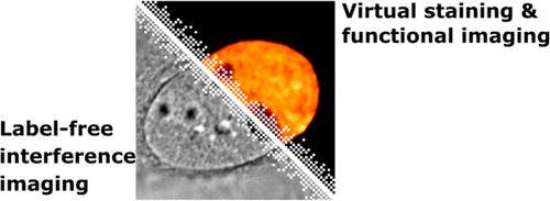
Molecularly Specific and Functional Live Cell Imaging by Label-Free Interference Microscopy
ACS Photonics ( IF 7 ) Pub Date : 2022-06-24 , DOI: 10.1021/acsphotonics.2c00599 Yi-Teng Hsiao, Chia-Ni Tsai, Ching-Ya Cheng, Chia-Lung Hsieh
ACS Photonics ( IF 7 ) Pub Date : 2022-06-24 , DOI: 10.1021/acsphotonics.2c00599 Yi-Teng Hsiao, Chia-Ni Tsai, Ching-Ya Cheng, Chia-Lung Hsieh
|
Optical interference microscopy is a powerful bioimaging technique by measuring the complex light fields associated with the specimen. Nowadays, the state-of-the-art interference microscopy makes it possible to directly visualize very small single biological nanoparticles and unlabeled macromolecules. The stable and indefinite linear scattering signal allows for continuous observation of the sample at a high speed, offering the opportunities to investigate single-molecule biophysics with the unprecedented details. Meanwhile, using interference microscopy to explore complex biological samples, such as a biological cell, emerges as an exciting research field. In this Perspective, we share our views on the impacts of optical interference microscopy on live cell imaging. Strategies for discriminating the scattering signals from different cell organelles and biological macromolecules are presented. In particular, the dynamic optical signal of live cells contains rich temporal information that is useful for enhancing the molecular specificity and functional information in label-free cell imaging. Finally, the challenges in three-dimensional imaging and turbidity suppression are discussed.
中文翻译:

通过无标记干涉显微镜进行分子特异性和功能性活细胞成像
光学干涉显微镜是一种强大的生物成像技术,通过测量与标本相关的复杂光场。如今,最先进的干涉显微镜可以直接观察非常小的单个生物纳米颗粒和未标记的大分子。稳定且不确定的线性散射信号允许以高速连续观察样品,提供了以前所未有的细节研究单分子生物物理学的机会。同时,使用干涉显微镜来探索复杂的生物样本,例如生物细胞,成为一个令人兴奋的研究领域。在这个观点中,我们分享了我们对光学干涉显微镜对活细胞成像影响的看法。提出了区分来自不同细胞器和生物大分子的散射信号的策略。特别是,活细胞的动态光信号包含丰富的时间信息,可用于增强无标记细胞成像中的分子特异性和功能信息。最后,讨论了三维成像和浊度抑制方面的挑战。
更新日期:2022-06-24
中文翻译:
通过无标记干涉显微镜进行分子特异性和功能性活细胞成像
光学干涉显微镜是一种强大的生物成像技术,通过测量与标本相关的复杂光场。如今,最先进的干涉显微镜可以直接观察非常小的单个生物纳米颗粒和未标记的大分子。稳定且不确定的线性散射信号允许以高速连续观察样品,提供了以前所未有的细节研究单分子生物物理学的机会。同时,使用干涉显微镜来探索复杂的生物样本,例如生物细胞,成为一个令人兴奋的研究领域。在这个观点中,我们分享了我们对光学干涉显微镜对活细胞成像影响的看法。提出了区分来自不同细胞器和生物大分子的散射信号的策略。特别是,活细胞的动态光信号包含丰富的时间信息,可用于增强无标记细胞成像中的分子特异性和功能信息。最后,讨论了三维成像和浊度抑制方面的挑战。


 京公网安备 11010802027423号