当前位置:
X-MOL 学术
›
Microsc. Res. Tech.
›
论文详情
Our official English website, www.x-mol.net, welcomes your feedback! (Note: you will need to create a separate account there.)
A phasor-based approach to improve optical sectioning in any confocal microscope with a tunable pinhole
Microscopy Research and Technique ( IF 2.5 ) Pub Date : 2022-06-10 , DOI: 10.1002/jemt.24178 Morgana D'Amico 1 , Elisabetta Di Franco 1 , Elena Cerutti 1, 2 , Vincenza Barresi 3 , Daniele Condorelli 3 , Alberto Diaspro 2, 4 , Luca Lanzanò 1, 2
Microscopy Research and Technique ( IF 2.5 ) Pub Date : 2022-06-10 , DOI: 10.1002/jemt.24178 Morgana D'Amico 1 , Elisabetta Di Franco 1 , Elena Cerutti 1, 2 , Vincenza Barresi 3 , Daniele Condorelli 3 , Alberto Diaspro 2, 4 , Luca Lanzanò 1, 2
Affiliation
|
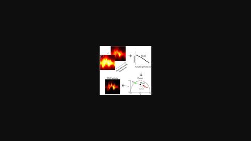
Confocal fluorescence microscopy is a well-established imaging technique capable of generating thin optical sections of biological specimens. Optical sectioning in confocal microscopy is mainly determined by the size of the pinhole, a small aperture placed in front of a point detector. In principle, imaging with a closed pinhole provides the highest degree of optical sectioning. In practice, the dramatic reduction of signal-to-noise ratio (SNR) at smaller pinhole sizes makes challenging the use of pinhole sizes significantly smaller than 1 Airy Unit (AU). Here, we introduce a simple method to “virtually” perform confocal imaging at smaller pinhole sizes without the dramatic reduction of SNR. The method is based on the sequential acquisition of multiple confocal images acquired at different pinhole aperture sizes and image processing based on a phasor analysis. The implementation is conceptually similar to separation of photons by lifetime tuning (SPLIT), a technique that exploits the phasor analysis to achieve super-resolution, and for this reason we call this method SPLIT-pinhole (SPLIT-PIN). We show with simulated data that the SPLIT-PIN image can provide improved optical sectioning (i.e., virtually smaller pinhole size) but better SNR with respect to an image obtained with closed pinhole. For instance, two images acquired at 2 and 1 AU can be combined to obtain a SPLIT-PIN image with a virtual pinhole size of 0.2 AU but with better SNR. As an example of application to biological imaging, we show that SPLIT-PIN improves confocal imaging of the apical membrane in an in vitro model of the intestinal epithelium.
中文翻译:

一种基于相量的方法,用于改进任何具有可调针孔的共聚焦显微镜中的光学切片
共聚焦荧光显微镜是一种成熟的成像技术,能够生成生物样本的薄光学切片。共聚焦显微镜中的光学切片主要由针孔的大小决定,针孔是放置在点检测器前面的小孔。原则上,使用闭合针孔成像可提供最高程度的光学切片。在实践中,较小的针孔尺寸会显着降低信噪比 (SNR),这使得使用明显小于 1 艾里单位 (AU) 的针孔尺寸具有挑战性。在这里,我们介绍了一种简单的方法,可以在较小的针孔尺寸下“虚拟”执行共焦成像,而不会显着降低 SNR。该方法基于在不同针孔孔径尺寸和基于相量分析的图像处理的多个共焦图像的顺序采集。该实现在概念上类似于通过寿命调谐 (SPLIT) 分离光子,这是一种利用相量分析来实现超分辨率的技术,因此我们将此方法称为 SPLIT-针孔 (SPLIT-PIN)。我们用模拟数据表明,SPLIT-PIN 图像可以提供改进的光学切片(即实际上更小的针孔尺寸),但相对于使用闭合针孔获得的图像而言,SNR 更好。例如,可以组合在 2 和 1 AU 处获取的两个图像以获得虚拟针孔大小为 0.2 AU 但具有更好 SNR 的 SPLIT-PIN 图像。作为生物成像应用的一个例子,
更新日期:2022-06-10
中文翻译:
一种基于相量的方法,用于改进任何具有可调针孔的共聚焦显微镜中的光学切片
共聚焦荧光显微镜是一种成熟的成像技术,能够生成生物样本的薄光学切片。共聚焦显微镜中的光学切片主要由针孔的大小决定,针孔是放置在点检测器前面的小孔。原则上,使用闭合针孔成像可提供最高程度的光学切片。在实践中,较小的针孔尺寸会显着降低信噪比 (SNR),这使得使用明显小于 1 艾里单位 (AU) 的针孔尺寸具有挑战性。在这里,我们介绍了一种简单的方法,可以在较小的针孔尺寸下“虚拟”执行共焦成像,而不会显着降低 SNR。该方法基于在不同针孔孔径尺寸和基于相量分析的图像处理的多个共焦图像的顺序采集。该实现在概念上类似于通过寿命调谐 (SPLIT) 分离光子,这是一种利用相量分析来实现超分辨率的技术,因此我们将此方法称为 SPLIT-针孔 (SPLIT-PIN)。我们用模拟数据表明,SPLIT-PIN 图像可以提供改进的光学切片(即实际上更小的针孔尺寸),但相对于使用闭合针孔获得的图像而言,SNR 更好。例如,可以组合在 2 和 1 AU 处获取的两个图像以获得虚拟针孔大小为 0.2 AU 但具有更好 SNR 的 SPLIT-PIN 图像。作为生物成像应用的一个例子,


 京公网安备 11010802027423号