Medical Image Analysis ( IF 10.9 ) Pub Date : 2022-05-25 , DOI: 10.1016/j.media.2022.102491 Mehdi Astaraki 1 , Örjan Smedby 2 , Chunliang Wang 2
|
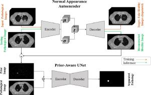
Segmentation of lung pathology in Computed Tomography (CT) images is of great importance for lung disease screening. However, the presence of different types of lung pathologies with a wide range of heterogeneities in size, shape, location, and texture, on one side, and their visual similarity with respect to surrounding tissues, on the other side, make it challenging to perform reliable automatic lesion segmentation. To leverage segmentation performance, we propose a deep learning framework comprising a Normal Appearance Autoencoder (NAA) model to learn the distribution of healthy lung regions and reconstruct pathology-free images from the corresponding pathological inputs by replacing the pathological regions with the characteristics of healthy tissues. Detected regions that represent prior information regarding the shape and location of pathologies are then integrated into a segmentation network to guide the attention of the model into more meaningful delineations. The proposed pipeline was tested on three types of lung pathologies, including pulmonary nodules, Non-Small Cell Lung Cancer (NSCLC), and Covid-19 lesion on five comprehensive datasets. The results show the superiority of the proposed prior model, which outperformed the baseline segmentation models in all the cases with significant margins. On average, adding the prior model improved the Dice coefficient for the segmentation of lung nodules by 0.038, NSCLCs by 0.101, and Covid-19 lesions by 0.041. We conclude that the proposed NAA model produces reliable prior knowledge regarding the lung pathologies, and integrating such knowledge into a prior segmentation network leads to more accurate delineations.
中文翻译:
用于肺病理学分割的先验自动编码器
计算机断层扫描 (CT) 图像中肺部病理的分割对于肺部疾病筛查具有重要意义。然而,一方面存在不同类型的肺部病变,在大小、形状、位置和质地方面存在广泛的异质性,另一方面,它们与周围组织的视觉相似性使得执行起来具有挑战性可靠的自动病灶分割。为了利用分割性能,我们提出了一个包含正常外观自动编码器(NAA)模型的深度学习框架,以学习健康肺区域的分布,并通过用健康组织的特征替换病理区域,从相应的病理输入重建无病理图像. 然后将检测到的表示有关病理形状和位置的先验信息的区域集成到分割网络中,以将模型的注意力引导到更有意义的描绘中。拟议的管道在五个综合数据集上对三种类型的肺部病变进行了测试,包括肺结节、非小细胞肺癌 (NSCLC) 和 Covid-19 病变。结果显示了所提出的先验模型的优越性,它在所有具有显着边际的情况下都优于基线分割模型。平均而言,添加先前模型将肺结节分割的 Dice 系数提高了 0.038,NSCLC 提高了 0.101,Covid-19 病灶提高了 0.041。我们得出结论,所提出的 NAA 模型产生了关于肺部病理学的可靠先验知识,


 京公网安备 11010802027423号