Acta Neuropathologica ( IF 12.7 ) Pub Date : 2022-05-17 , DOI: 10.1007/s00401-022-02427-2 Nicole L Ackermans 1, 2, 3, 4 , Merina Varghese 1, 2 , Terrie M Williams 5 , Nicholas Grimaldi 1, 2 , Enna Selmanovic 1, 2 , Akbar Alipour 2, 6 , Priti Balchandani 2, 6 , Joy S Reidenberg 3 , Patrick R Hof 1, 2, 7
|
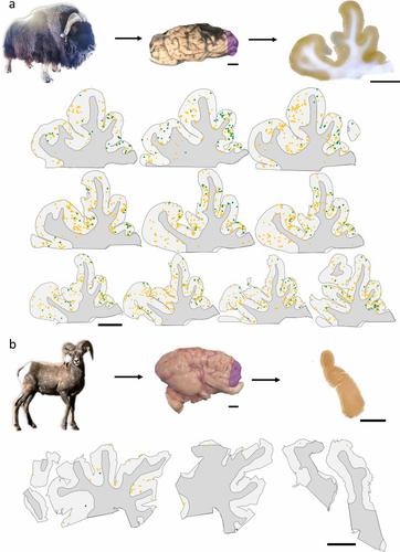
Traumatic brain injury (TBI) is a leading cause of neurologic impairment and death that remains poorly understood. Rodent models have yet to produce clinical therapies, and the exploration of larger and more diverse models remains relatively scarce. We investigated the potential for brain injury after headbutting in two combative bovid species by assessing neuromorphology and neuropathology through immunohistochemistry and stereological quantification. Postmortem brains of muskoxen (Ovibos moschatus, n = 3) and bighorn sheep (Ovis canadensis, n = 4) were analyzed by high-resolution MRI and processed histologically for evidence of TBI. Exploratory histological protocols investigated potential abnormalities in neurons, microglia, and astrocytes in the prefrontal and parietal cortex. Phosphorylated tau protein, a TBI biomarker found in the cerebrospinal fluid and in neurodegenerative lesions, was used to detect possible cellular consequences of chronic or acute TBI. MRI revealed no abnormal neuropathological changes; however, high amounts of tau-immunoreactive neuritic thread clusters, neurites, and neurons were concentrated in the superficial layers of the neocortex, preferentially at the bottom of the sulci in the muskoxen and occasionally around blood vessels. Tau-immunoreactive lesions were rare in the bighorn sheep. Additionally, microglia and astrocytes showed no grouping around tau-immunoreactive cells in either species. Our preliminary findings indicate that muskoxen and possibly other headbutting bovids suffer from chronic or acute brain trauma and that the males’ thicker skulls may protect them to a certain extent.
中文翻译:
撞头牛科创伤性脑损伤的证据
创伤性脑损伤 (TBI) 是导致神经功能障碍和死亡的主要原因,但目前仍知之甚少。啮齿动物模型尚未产生临床疗法,对更大、更多样化模型的探索仍然相对稀缺。我们通过免疫组织化学和体视学量化评估神经形态学和神经病理学,研究了两种好斗牛科动物头部撞击后脑损伤的可能性。麝牛 ( Ovibos moschatus , n = 3) 和大角羊 ( Ovis canadensis , n的死后大脑 = 4) 通过高分辨率 MRI 进行分析,并在组织学上处理 TBI 的证据。探索性组织学方案研究了前额叶和顶叶皮层中神经元、小胶质细胞和星形胶质细胞的潜在异常。磷酸化 tau 蛋白是一种在脑脊液和神经退行性病变中发现的 TBI 生物标志物,用于检测慢性或急性 TBI 可能的细胞后果。MRI未发现异常的神经病理学变化;然而,大量的 tau 免疫反应性神经炎线簇、神经突和神经元集中在新皮层的表层,优先位于 muskoxen 的脑沟底部,偶尔在血管周围。Tau 免疫反应性病变在大角羊中很少见。此外,小胶质细胞和星形胶质细胞在这两个物种中都没有显示出围绕 tau 免疫反应细胞的分组。我们的初步研究结果表明,麝香牛和可能的其他撞头牛患有慢性或急性脑外伤,雄性较厚的头骨可以在一定程度上保护它们。


 京公网安备 11010802027423号