当前位置:
X-MOL 学术
›
Chem. Soc. Rev.
›
论文详情
Our official English website, www.x-mol.net, welcomes your feedback! (Note: you will need to create a separate account there.)
Photo-induced force microscopy (PiFM) – principles and implementations
Chemical Society Reviews ( IF 46.2 ) Pub Date : 2022-05-05 , DOI: 10.1039/d2cs00052k Abid Anjum Sifat 1 , Junghoon Jahng 2 , Eric O Potma 1, 3
Chemical Society Reviews ( IF 46.2 ) Pub Date : 2022-05-05 , DOI: 10.1039/d2cs00052k Abid Anjum Sifat 1 , Junghoon Jahng 2 , Eric O Potma 1, 3
Affiliation
|
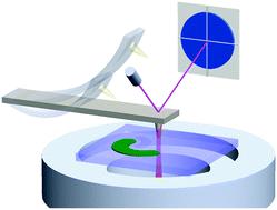
Photo-induced force microscopy (PiFM) is a scan probe technique that offers images with spectroscopic contrast at a spatial resolution in the nanometer range. PiFM utilizes the non-propagating, enhanced near field at the apex of a sharp tip to locally induce a polarization in the sample, which in turn produces an additional force acting on the cantilevered tip. This photo-induced force, though in the pN range or less, can be extracted from the oscillation properties of the cantilever, thus enabling the generation of photo-induced force maps. Since its inception in 2010, the PiFM technique has grown into a useful nano-spectrocopic tool that has expanded its reach in terms of imaging capabilities and applications. In this review, we present various technical implementations of the PiFM approach. In addition, we discuss the physical origin of the PiFM signal, highlighting the contributions from dipole–dipole forces as well as forces that derive from photo-thermal processes.
中文翻译:

光致力显微镜 (PiFM) – 原理和实现
光致力显微镜 (PiFM) 是一种扫描探针技术,可在纳米范围内的空间分辨率下提供具有光谱对比度的图像。PiFM 利用尖锐尖端顶点处的非传播增强近场在样品中局部诱导极化,从而产生作用在悬臂尖端上的附加力。这种光致力虽然在 pN 范围内或更小,但可以从悬臂的振荡特性中提取,从而能够生成光致力图。自 2010 年问世以来,PiFM 技术已发展成为一种有用的纳米分光镜工具,在成像能力和应用方面扩大了其范围。在这篇评论中,我们介绍了 PiFM 方法的各种技术实现。此外,
更新日期:2022-05-05
中文翻译:
光致力显微镜 (PiFM) – 原理和实现
光致力显微镜 (PiFM) 是一种扫描探针技术,可在纳米范围内的空间分辨率下提供具有光谱对比度的图像。PiFM 利用尖锐尖端顶点处的非传播增强近场在样品中局部诱导极化,从而产生作用在悬臂尖端上的附加力。这种光致力虽然在 pN 范围内或更小,但可以从悬臂的振荡特性中提取,从而能够生成光致力图。自 2010 年问世以来,PiFM 技术已发展成为一种有用的纳米分光镜工具,在成像能力和应用方面扩大了其范围。在这篇评论中,我们介绍了 PiFM 方法的各种技术实现。此外,


 京公网安备 11010802027423号