GeroScience ( IF 5.6 ) Pub Date : 2022-04-28 , DOI: 10.1007/s11357-022-00576-6 Chao-Wen Lin, Tzu-Ting Lai, Szu-Ju Chen, Chin-Hsien Lin
|
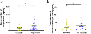
The pathognomonic hallmark of Parkinson’s disease (PD), α-synuclein, has been observed in the retina of PD patients. We investigated whether biomarkers in the tears and retinal microvascular changes associate with PD risk and progression. This prospective study enrolled 49 PD patients and 45 age-matched healthy controls. The α-synuclein and neurofilament light chain (NfL) levels were measured using an electrochemiluminescence immunoassay. Retinal vessel density was assessed using optical coherence tomography angiography (OCT-A). The Movement Disorder Society-Unified Parkinson’s Disease Rating Scale (MDS-UPDRS) and Mini-Mental State Examination score were used to assess motor and cognitive progression. The α-synuclein and NfL levels in the tears were higher in PD patients than in controls (α-synuclein: 55.49 ± 8.12 pg/mL vs. 31.71 ± 3.25 pg/mL, P = 0.009; NfL: 2.89 ± 0.52 pg/mL vs. 1.47 ± 0.23 pg/mL, P = 0.02). The vessel densities in the deep plexus of central macula and the radial peripapillary capillary layer of disc region were lower in PD patients with moderate-stage compared with early-stage PD (P < 0.05). The accuracy of predicting PD occurrence using age and sex alone (area under the curve [AUC] 0.612) was significantly improved by adding α-synuclein and NfL levels and retinal vascular densities (AUC 0.752, P = 0.001). After a mean follow-up of 1.5 ± 0.3 years, the accuracy of predicting motor or cognitive progression using age, sex, and baseline motor severity as a basic model was increased by incorporating retinal microvascular and biofluid markers as a full model (P = 0.001). Our results showed that retinal microvascular densities combined with α-synuclein and NfL levels in tears are associated with risk and progression of PD.
中文翻译:
帕金森病患者泪液中 α-突触核蛋白和 NfL 水平升高,视网膜微血管密度降低
已在 PD 患者的视网膜中观察到帕金森病 (PD) 的典型特征 α-突触核蛋白。我们研究了眼泪和视网膜微血管变化中的生物标志物是否与 PD 风险和进展相关。这项前瞻性研究招募了 49 名 PD 患者和 45 名年龄匹配的健康对照。使用电化学发光免疫测定法测量 α-突触核蛋白和神经丝轻链 (NfL) 水平。使用光学相干断层扫描血管造影术(OCT-A)评估视网膜血管密度。运动障碍协会-统一帕金森病评定量表 (MDS-UPDRS) 和简易精神状态检查评分用于评估运动和认知进展。PD 患者泪液中的 α-突触核蛋白和 NfL 水平高于对照组(α-突触核蛋白:55.49 ± 8.12 pg/mL vs. 31.71 ± 3.25 pg/mL,P = 0.009;NfL:2.89 ± 0.52 pg/mL 与 1.47 ± 0.23 pg/mL,P = 0.02)。与早期PD相比,中期PD患者中央黄斑深部神经丛和椎间盘区径向毛细血管层的血管密度较低(P < 0.05)。通过添加 α-突触核蛋白和 NfL 水平以及视网膜血管密度(AUC 0.752,P = 0.001),仅使用年龄和性别(曲线下面积 [AUC] 0.612)预测 PD 发生的准确性显着提高。在 1.5 ± 0.3 年的平均随访后,通过将视网膜微血管和生物流体标记物作为完整模型,使用年龄、性别和基线运动严重程度作为基本模型预测运动或认知进展的准确性提高了。P = 0.001)。我们的研究结果表明,泪液中视网膜微血管密度与 α-突触核蛋白和 NfL 水平相结合,与 PD 的风险和进展有关。


 京公网安备 11010802027423号