Our official English website, www.x-mol.net, welcomes your feedback! (Note: you will need to create a separate account there.)
Single-Molecule Micromanipulation and Super-Resolution Imaging Resolve Nanodomains Underlying Chromatin Folding in Mitotic Chromosomes
ACS Nano ( IF 17.1 ) Pub Date : 2022-04-29 , DOI: 10.1021/acsnano.2c01025 Jiabin Wang 1 , Chuansheng Hu 2 , Xuecheng Chen 1 , Yaqian Li 2 , Jielin Sun 1 , Daniel M Czajkowsky 2 , Zhifeng Shao 2
ACS Nano ( IF 17.1 ) Pub Date : 2022-04-29 , DOI: 10.1021/acsnano.2c01025 Jiabin Wang 1 , Chuansheng Hu 2 , Xuecheng Chen 1 , Yaqian Li 2 , Jielin Sun 1 , Daniel M Czajkowsky 2 , Zhifeng Shao 2
Affiliation
|
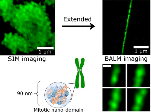
The folding of interphase chromatin into highly compact mitotic chromosomes is one of the most recognizable changes during the cell cycle. However, the structural organization underlying this drastic compaction remains elusive. Here, we combine several super resolution methods, including structured illumination microscopy (SIM), binding-activated localization microscopy (BALM), and atomic force microscopy (AFM), to examine the structural details of the DNA within the mitotic chromosome, both in the native state and after up to 30-fold extension using single-molecule micromanipulation. Images of native chromosomes reveal widespread ∼125 nm compact granules (CGs) throughout the metaphase chromosome. However, at maximal extensions, we find exclusively ∼90 nm domains (mitotic nanodomains, MNDs) that are unexpectedly resistant to extensive forces of tens of nanonewtons. The DNA content of the MNDs is estimated to be predominantly ∼80 kb, which is comparable to the size of the inner loops predicted by a recent nested loop model of the mitotic chromosome. With this DNA content, the total volume expected of the human genome assuming closely packed MNDs is nearly identical to what is observed. Thus, altogether, these results suggest that these mechanically stable MNDs, and their higher-order assembly into CGs, are the dominant higher-level structures that underlie the compaction of chromatin from interphase to metaphase.
中文翻译:

单分子显微操作和超分辨率成像解决有丝分裂染色体中染色质折叠的纳米结构域
间期染色质折叠成高度紧凑的有丝分裂染色体是细胞周期中最明显的变化之一。然而,这种剧烈压实背后的结构组织仍然难以捉摸。在这里,我们结合了几种超分辨率方法,包括结构照明显微镜 (SIM)、结合激活定位显微镜 (BALM) 和原子力显微镜 (AFM),以检查有丝分裂染色体内 DNA 的结构细节,无论是在天然状态和使用单分子显微操作进行高达 30 倍的扩展后。原生染色体的图像揭示了整个中期染色体广泛分布的 ∼125 nm 致密颗粒 (CG)。然而,在最大延伸处,我们发现只有 ∼90 nm 域(有丝分裂纳米域,MNDs)出乎意料地抵抗了数十纳牛顿的广泛力量。据估计,MND 的 DNA 含量主要为 ~80 kb,这与最近的有丝分裂染色体嵌套环模型预测的内环大小相当。有了这个 DNA 含量,假设紧密堆积的 MND,人类基因组的预期总体积与观察到的几乎相同。因此,总的来说,这些结果表明,这些机械稳定的 MND 及其高阶组装成 CG,是染色质从间期到中期压实的主要高级结构。有了这个 DNA 含量,假设紧密堆积的 MND,人类基因组的预期总体积与观察到的几乎相同。因此,总的来说,这些结果表明,这些机械稳定的 MND 及其高阶组装成 CG,是染色质从间期到中期压实的主要高级结构。有了这个 DNA 含量,假设紧密堆积的 MND,人类基因组的预期总体积与观察到的几乎相同。因此,总的来说,这些结果表明,这些机械稳定的 MND 及其高阶组装成 CG,是染色质从间期到中期压实的主要高级结构。
更新日期:2022-04-29
中文翻译:
单分子显微操作和超分辨率成像解决有丝分裂染色体中染色质折叠的纳米结构域
间期染色质折叠成高度紧凑的有丝分裂染色体是细胞周期中最明显的变化之一。然而,这种剧烈压实背后的结构组织仍然难以捉摸。在这里,我们结合了几种超分辨率方法,包括结构照明显微镜 (SIM)、结合激活定位显微镜 (BALM) 和原子力显微镜 (AFM),以检查有丝分裂染色体内 DNA 的结构细节,无论是在天然状态和使用单分子显微操作进行高达 30 倍的扩展后。原生染色体的图像揭示了整个中期染色体广泛分布的 ∼125 nm 致密颗粒 (CG)。然而,在最大延伸处,我们发现只有 ∼90 nm 域(有丝分裂纳米域,MNDs)出乎意料地抵抗了数十纳牛顿的广泛力量。据估计,MND 的 DNA 含量主要为 ~80 kb,这与最近的有丝分裂染色体嵌套环模型预测的内环大小相当。有了这个 DNA 含量,假设紧密堆积的 MND,人类基因组的预期总体积与观察到的几乎相同。因此,总的来说,这些结果表明,这些机械稳定的 MND 及其高阶组装成 CG,是染色质从间期到中期压实的主要高级结构。有了这个 DNA 含量,假设紧密堆积的 MND,人类基因组的预期总体积与观察到的几乎相同。因此,总的来说,这些结果表明,这些机械稳定的 MND 及其高阶组装成 CG,是染色质从间期到中期压实的主要高级结构。有了这个 DNA 含量,假设紧密堆积的 MND,人类基因组的预期总体积与观察到的几乎相同。因此,总的来说,这些结果表明,这些机械稳定的 MND 及其高阶组装成 CG,是染色质从间期到中期压实的主要高级结构。


 京公网安备 11010802027423号