Our official English website, www.x-mol.net, welcomes your feedback! (Note: you will need to create a separate account there.)
Micro-/nano-fluidic devices and in vivo fluorescence imaging based on quantum dots for cytologic diagnosis
Lab on a Chip ( IF 6.1 ) Pub Date : 2022-04-22 , DOI: 10.1039/d2lc00113f Minchuan Luo 1 , Hiroshi Yukawa 1, 2, 3, 4, 5 , Yoshinobu Baba 1, 2, 3
Lab on a Chip ( IF 6.1 ) Pub Date : 2022-04-22 , DOI: 10.1039/d2lc00113f Minchuan Luo 1 , Hiroshi Yukawa 1, 2, 3, 4, 5 , Yoshinobu Baba 1, 2, 3
Affiliation
|
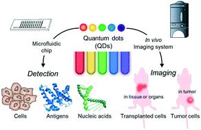
Semiconductor quantum dots (QDs) possess attractive merits over traditional organic dyes, such as tunable emission, narrow emission spectra and good resistance against optical bleaching, and play a vital role in biosensing and bioimaging for cytologic diagnoses. Microfluidic technology is a potentially useful strategy, as it provides a rapid platform for tracing of disease markers. In vivo fluorescence imaging (FI) based on QDs has become popular for the analysis of complex biological processes. We herein report the applications of multifunctional fluorescent QDs as sensitive probes for diagnoses on cancer medicine and stem cell therapy via microfluidic chips and in vivo imaging.
中文翻译:

用于细胞学诊断的基于量子点的微/纳流体装置和体内荧光成像
与传统有机染料相比,半导体量子点(QD)具有可调谐发射、窄发射光谱和良好的抗光学漂白性等优点,在细胞学诊断的生物传感和生物成像中发挥着至关重要的作用。微流体技术是一种潜在有用的策略,因为它为追踪疾病标志物提供了一个快速平台。基于量子点的体内荧光成像 (FI) 已成为分析复杂生物过程的流行方法。我们在此报告了多功能荧光量子点作为敏感探针通过微流控芯片和体内成像诊断癌症医学和干细胞治疗的应用。
更新日期:2022-04-22
中文翻译:
用于细胞学诊断的基于量子点的微/纳流体装置和体内荧光成像
与传统有机染料相比,半导体量子点(QD)具有可调谐发射、窄发射光谱和良好的抗光学漂白性等优点,在细胞学诊断的生物传感和生物成像中发挥着至关重要的作用。微流体技术是一种潜在有用的策略,因为它为追踪疾病标志物提供了一个快速平台。基于量子点的体内荧光成像 (FI) 已成为分析复杂生物过程的流行方法。我们在此报告了多功能荧光量子点作为敏感探针通过微流控芯片和体内成像诊断癌症医学和干细胞治疗的应用。


 京公网安备 11010802027423号