Progress in Retinal and Eye Research ( IF 17.8 ) Pub Date : 2022-01-21 , DOI: 10.1016/j.preteyeres.2021.101038 Imran H Yusuf 1 , Andrew M Garrett 2 , Robert E MacLaren 1 , Peter Charbel Issa 1
|
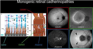
Cadherins are a superfamily of calcium-dependent intercellular adhesion molecules that are widely expressed in living tissues. Within the retina and retinal pigment epithelium (RPE), cadherins contribute to tissue morphogenesis, neural circuit formation, adherens junctions of the outer blood-retinal barrier, photoreceptor disc morphogenesis, maintenance and survival. Four monogenic disorders involving genes which encode cadherins have been identified as causes of inherited retinal degeneration: the retinal cadherinopathies (CDHR1, CDH23, PCDH15, CDH3). Biallelic variants in CDHR1 result in cone-rod dystrophy, rod-cone dystrophy or late-onset macular dystrophy which may be misclassified as dry age-related macular degeneration. Biallelic variants in CDH23 and PCDH15 underlie Usher Syndrome type 1D and 1F. Hypotrichosis with juvenile macular dystrophy results from biallelic variants in CDH3, which contributes to adherens tight junctions between RPE cells. In this review, we summarise the classification of cadherins, and the role of cadherins in the physiology and morphogenesis of the inner and outer retina. Cadherins expressed in primate photoreceptors (CDHR1, CDH23 and PCDH15) have evolved complex roles in outer segment disc morphogenesis and maintenance involving intracellular heterophilic interactions which are as yet incompletely characterised. We highlight what is currently unknown about the molecular function of these cadherins, and review the pathogenesis, clinical phenotype and molecular genetics of each monogenic retinal cadherinopathy. Genes regulating the expression and post-translational modification of retinal cadherins, or those coding for as yet unidentified interacting partners, are candidates for unsolved cases of retinal degeneration. This group of disorders is potentially treatable; we summarise the likely molecular therapeutic approaches and future directions for each retinal cadherinopathy.
中文翻译:
视网膜钙粘蛋白和视网膜钙粘蛋白病:当前概念和未来方向
钙粘蛋白是钙依赖性细胞间粘附分子的超家族,在活组织中广泛表达。在视网膜和视网膜色素上皮 (RPE) 内,钙粘蛋白有助于组织形态发生、神经回路形成、外层血-视网膜屏障的粘附连接、感光盘形态发生、维持和存活。四种涉及编码钙粘蛋白基因的单基因疾病已被确定为遗传性视网膜变性的原因:视网膜钙粘蛋白病(CDHR1、CDH23、PCDH15、CDH3)。CDHR1 中的双等位基因变异会导致视杆细胞营养不良、视杆视锥细胞营养不良或迟发性黄斑营养不良,这可能被错误归类为干性年龄相关性黄斑变性。CDH23 中的双等位基因变体和PCDH15是 Usher 综合征 1D 和 1F 型的基础。CDH3双等位基因变异导致幼年型黄斑营养不良的少毛症,这有助于 RPE 细胞之间的粘附紧密连接。在这篇综述中,我们总结了钙粘蛋白的分类,以及钙粘蛋白在内外视网膜的生理和形态发生中的作用。在灵长类动物光感受器(CDHR1、CDH23 和 PCDH15)中表达的钙粘蛋白在外段椎间盘形态发生和维持中发挥了复杂的作用,涉及尚未完全表征的细胞内嗜异性相互作用。我们重点介绍了这些钙粘蛋白分子功能目前未知的内容,并回顾了每种单基因视网膜钙粘蛋白病的发病机制、临床表型和分子遗传学。调节视网膜钙粘蛋白表达和翻译后修饰的基因,或编码尚未确定的相互作用伙伴的基因,是未解决的视网膜变性病例的候选者。这组疾病是可以治疗的;我们总结了每种视网膜钙粘蛋白病可能的分子治疗方法和未来方向。


 京公网安备 11010802027423号