Medical Image Analysis ( IF 10.9 ) Pub Date : 2021-10-29 , DOI: 10.1016/j.media.2021.102269 Shunli Wang 1 , François Varray 2 , Wanyu Liu 3 , Patrick Clarysse 2 , Isabelle E Magnin 2
|
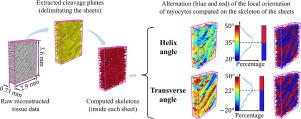
Most cardiomyocytes in the left ventricle wall are grouped in aggregates of four to five units that are quasi-parallel to each other. When one or more “cardiomyocyte aggregates” are delimited by two cleavage planes, this defines a “sheetlet” that can be considered as a “work unit” that contributes to the thickening of the wall during the cardiac cycle. In this paper, we introduce the skeleton method to measure the local three-dimensional (3D) orientation of cardiomyocyte aggregates in the sheetlets in three steps: data segmentation; extraction of the skeleton of the sheetlets; and calculation of the local orientation of the cardiomyocyte aggregates inside the sheetlets. These data include a series of virtual tissue volumes and five transmural human left ventricle free wall samples, imaged with 3D synchrotron radiation phase-contrast microtomography, and reconstructed with a voxel size. We computed the local orientation of the cardiomyocyte aggregates inside the sheetlets with a working window of in size. These data demonstrate that the skeleton method can provide accurate 3D measurements and reliable screening of the 3D evolution of the orientation of cardiomyocyte aggregates within the sheetlets. We showed that in regions that contain one population of quasi-parallel sheetlets, the orientation of the cardiomyocyte aggregates undergo “oscillations” along the perpendicular direction of the sheetlets. In regions that contain two populations of sheetlets with a different angular range, we demonstrate some discontinuity of the helix angle of the cardiomyocyte aggregates at the interface between the two populations.
中文翻译:
使用 X 射线相差显微断层扫描测量人左心室游离壁样品中心肌细胞聚集体的局部方向
左心室壁中的大多数心肌细胞聚集成四到五个彼此准平行的单元。当一个或多个“心肌细胞聚集体”由两个分裂平面界定时,这定义了一个“薄片”,可被视为一个“工作单元”,有助于在心动周期中使壁增厚。在本文中,我们介绍了骨架方法分三个步骤测量薄片中心肌细胞聚集体的局部三维 (3D) 方向:数据分割;提取薄片的骨架;并计算薄片内心肌细胞聚集体的局部方向。这些数据包括一系列虚拟组织体积和五个透壁人体左心室游离壁样本,用 3D 同步辐射相衬显微断层扫描成像,并用 体素大小。我们计算了薄片内心肌细胞聚集体的局部方向,工作窗口为在尺寸方面。这些数据表明,骨架方法可以提供准确的 3D 测量和可靠筛选薄片内心肌细胞聚集体方向的 3D 演变。我们发现,在包含一组准平行薄片的区域中,心肌细胞聚集体的方向沿着薄片的垂直方向发生“振荡”。在包含两个具有不同角度范围的薄片群体的区域中,我们证明了在两个群体之间的界面处心肌细胞聚集体的螺旋角存在一些不连续性。


 京公网安备 11010802027423号