Applied Surface Science ( IF 6.7 ) Pub Date : 2021-09-24 , DOI: 10.1016/j.apsusc.2021.151310 Elvis O. López 1 , Pablo L. Bernardo 2 , Noemi R. Checca 1 , André L. Rossi 1 , Alexandre Mello 1 , Donald E. Ellis 3 , Alexandre M. Rossi 1 , Joice Terra 1
|
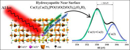
X-ray photoelectron spectroscopy (XPS) is one of the main tools for hydroxyapatite (HA) surface characterization in developing materials for biomedical and heterogeneous catalysis. Despite the XPS technique's potential to correlate binding energies with existing photo-emitter sites on near-surfaces, few previous studies analyzed this aspect for HA and metal-substituted HA surfaces. In this work, we theoretically reconstructed the XPS spectra of stoichiometric HA and lead-substituted hydroxyapatite (PbCaHA, Ca10-xPbx(PO4)6(OH)2; x = 2, 10) using a first-principles linear combination of atomic orbitals embedded cluster approach and periodic supercell band structures within the framework of Density Functional Theory (DFT). We take into account photoemission lines contributions from Ca(1), Ca(2), Pb(1) and Pb(2) sites located on surface and near-surface depths (up to ∼15 Å) along the (0 0 1) and (1 0 0) surfaces. The calculated DFT spectra of HA and PbCaHA were compared with high-resolution XPS spectra previously characterized by synchrotron X-ray diffraction (XRD), Fourier transforms infrared spectroscopy (FTIR), high-resolution transmission electron microscopy (HRTEM) and electron energy-loss spectroscopy (EELS). A combined theoretical and experimental approach enables decoding of the complex structure of HA and PbCaHA in XPS spectra. It was found that XPS binding energies profiles depend significantly on photo-emitters from near-surface sites and surface crystallographic orientation. The main Ca 2p3/2 envelope peak in HA is predominantly from Ca(1) and Ca(2) sites (∼347.4 eV), while the weaker peak is due to the Ca(2) site only (∼345.0 eV). Variations on HA nanoparticle morphology could be a critical factor for changes in XPS binding energies' profile.
中文翻译:
羟基磷灰石和铅取代的羟基磷灰石近表面结构:来自 X 射线光电子光谱的光电发射线的新模型
X 射线光电子能谱 (XPS) 是羟基磷灰石 (HA) 表面表征的主要工具之一,用于开发生物医学和多相催化材料。尽管 XPS 技术有可能将结合能与近表面上现有的光发射器位点相关联,但以前的研究很少分析 HA 和金属取代的 HA 表面的这一方面。在这项工作中,我们从理论上重建了化学计量的 HA 和铅取代的羟基磷灰石 (PbCaHA, Ca 10-x Pb x (PO 4 ) 6 (OH) 2 )的 XPS 光谱; x = 2, 10) 在密度泛函理论 (DFT) 的框架内使用原子轨道嵌入簇方法和周期性超胞带结构的第一性原理线性组合。我们考虑了沿 (0 0 1)位于表面和近表面深度(高达 15 Å)的 Ca(1)、Ca(2)、Pb(1) 和 Pb(2) 位点的光发射线贡献和 (1 0 0) 表面。HA 和 PbCaHA 的计算 DFT 光谱与先前通过同步加速器 X 射线衍射 (XRD)、傅里叶变换红外光谱 (FTIR)、高分辨率透射电子显微镜 (HRTEM) 和电子能量损失表征的高分辨率 XPS 光谱进行了比较光谱(鳗鱼)。理论和实验相结合的方法能够解码 XPS 光谱中 HA 和 PbCaHA 的复杂结构。发现 XPS 结合能分布显着取决于来自近表面位点的光发射器和表面晶体取向。主Ca 2p 3/2HA 中的包络峰主要来自 Ca(1) 和 Ca(2) 位点 (~347.4 eV),而较弱的峰仅来自 Ca(2) 位点 (~345.0 eV)。HA 纳米颗粒形态的变化可能是 XPS 结合能分布变化的关键因素。


 京公网安备 11010802027423号