当前位置:
X-MOL 学术
›
Anal. Chem.
›
论文详情
Our official English website, www.x-mol.net, welcomes your feedback! (Note: you will need to create a separate account there.)
Nanodissection of Selected Viral Particles by Scanning Transmission Electron Microscopy/Focused Ion Beam for Genetic Identification
Analytical Chemistry ( IF 7.4 ) Pub Date : 2021-09-22 , DOI: 10.1021/acs.analchem.1c01001 Dror Horvitz 1 , Elad Milrot 2 , Neta Luria 3 , Efi Makdasi 2 , Adi Beth-Din 4 , Itai Glinert 2 , Aviv Dombrovsky 3 , Orly Laskar 2
Analytical Chemistry ( IF 7.4 ) Pub Date : 2021-09-22 , DOI: 10.1021/acs.analchem.1c01001 Dror Horvitz 1 , Elad Milrot 2 , Neta Luria 3 , Efi Makdasi 2 , Adi Beth-Din 4 , Itai Glinert 2 , Aviv Dombrovsky 3 , Orly Laskar 2
Affiliation
|
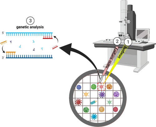
This study presents the development of a new correlative workflow to bridge the gap between electron microscopy imaging and genetic analysis of viruses. The workflow enables the assignment of genetic information to a specific biological entity by harnessing the nanodissection capability of focused ion beam (FIB). This correlative workflow is based on scanning transmission electron microscopy (STEM) and FIB followed by a polymerase chain reaction (PCR). For this purpose, we studied the tomato brown rugose fruit virus (ToBRFV) and the adenovirus that have significant impacts on plant integrity and human health, respectively. STEM imaging was used for the identification and localization of virus particles on a transmission electron microscopy (TEM) grid followed by FIB milling of the desired region of interest. The final-milled product was subjected to genetic analysis by the PCR. The results prove that the FIB-milling process maintains the integrity of the genetic material as confirmed by the PCR. We demonstrate the identification of RNA and DNA viruses extracted from a few micrometers of an FIB-milled TEM grid. This workflow enables the genetic analysis of specifically imaged viral particles directly from heterogeneous clinical samples. In addition to viral diagnostics, the ability to isolate and to genetically identify specific submicrometer structures may prove valuable in additional fields, including subcellular organelle and granule research.
中文翻译:

通过扫描透射电子显微镜/聚焦离子束对选定的病毒颗粒进行纳米切割以进行遗传鉴定
这项研究提出了一种新的相关工作流程的发展,以弥合电子显微镜成像和病毒遗传分析之间的差距。该工作流程通过利用聚焦离子束 (FIB) 的纳米切割能力将遗传信息分配给特定的生物实体。此相关工作流程基于扫描透射电子显微镜 (STEM) 和 FIB,然后是聚合酶链反应 (PCR)。为此,我们分别研究了对植物完整性和人类健康有显着影响的番茄褐色皱纹果实病毒 (ToBRFV) 和腺病毒。STEM 成像用于在透射电子显微镜 (TEM) 网格上识别和定位病毒颗粒,然后对所需的感兴趣区域进行 FIB 研磨。最终研磨的产品通过 PCR 进行遗传分析。结果证明 FIB 研磨过程保持了 PCR 所证实的遗传材料的完整性。我们展示了从几微米的 FIB 研磨 TEM 网格中提取的 RNA 和 DNA 病毒的鉴定。该工作流程可以直接从异质临床样本中对特定成像的病毒颗粒进行遗传分析。除了病毒诊断外,分离和遗传识别特定亚微米结构的能力可能在其他领域(包括亚细胞器和颗粒研究)中证明是有价值的。我们展示了从几微米的 FIB 研磨 TEM 网格中提取的 RNA 和 DNA 病毒的鉴定。该工作流程可以直接从异质临床样本中对特定成像的病毒颗粒进行遗传分析。除了病毒诊断外,分离和遗传识别特定亚微米结构的能力可能在其他领域(包括亚细胞器和颗粒研究)中证明是有价值的。我们展示了从几微米的 FIB 研磨 TEM 网格中提取的 RNA 和 DNA 病毒的鉴定。该工作流程可以直接从异质临床样本中对特定成像的病毒颗粒进行遗传分析。除了病毒诊断外,分离和遗传识别特定亚微米结构的能力可能在其他领域(包括亚细胞器和颗粒研究)中证明是有价值的。
更新日期:2021-10-06
中文翻译:
通过扫描透射电子显微镜/聚焦离子束对选定的病毒颗粒进行纳米切割以进行遗传鉴定
这项研究提出了一种新的相关工作流程的发展,以弥合电子显微镜成像和病毒遗传分析之间的差距。该工作流程通过利用聚焦离子束 (FIB) 的纳米切割能力将遗传信息分配给特定的生物实体。此相关工作流程基于扫描透射电子显微镜 (STEM) 和 FIB,然后是聚合酶链反应 (PCR)。为此,我们分别研究了对植物完整性和人类健康有显着影响的番茄褐色皱纹果实病毒 (ToBRFV) 和腺病毒。STEM 成像用于在透射电子显微镜 (TEM) 网格上识别和定位病毒颗粒,然后对所需的感兴趣区域进行 FIB 研磨。最终研磨的产品通过 PCR 进行遗传分析。结果证明 FIB 研磨过程保持了 PCR 所证实的遗传材料的完整性。我们展示了从几微米的 FIB 研磨 TEM 网格中提取的 RNA 和 DNA 病毒的鉴定。该工作流程可以直接从异质临床样本中对特定成像的病毒颗粒进行遗传分析。除了病毒诊断外,分离和遗传识别特定亚微米结构的能力可能在其他领域(包括亚细胞器和颗粒研究)中证明是有价值的。我们展示了从几微米的 FIB 研磨 TEM 网格中提取的 RNA 和 DNA 病毒的鉴定。该工作流程可以直接从异质临床样本中对特定成像的病毒颗粒进行遗传分析。除了病毒诊断外,分离和遗传识别特定亚微米结构的能力可能在其他领域(包括亚细胞器和颗粒研究)中证明是有价值的。我们展示了从几微米的 FIB 研磨 TEM 网格中提取的 RNA 和 DNA 病毒的鉴定。该工作流程可以直接从异质临床样本中对特定成像的病毒颗粒进行遗传分析。除了病毒诊断外,分离和遗传识别特定亚微米结构的能力可能在其他领域(包括亚细胞器和颗粒研究)中证明是有价值的。


 京公网安备 11010802027423号