Annals of Anatomy ( IF 2.2 ) Pub Date : 2021-09-23 , DOI: 10.1016/j.aanat.2021.151835 Harry Etienne 1 , Jésus Gonzalez-Bermejo 2 , Martin Dres 3 , Thierry Maisonobe 4 , Guy Brochier 4 , Laure Wingertsmann 5 , Olivier Thibaudeau 5 , Hicham Masmoudi 6 , Jalal Assouad 1 , Thomas Similowski 7
|
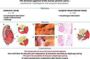
Background
Diaphragm pacing allows certain ventilator-dependent patients to achieve weaning from mechanical ventilation. The reference method consists in implanting intrathoracic contact electrodes around the phrenic nerve during video-assisted thoracic surgery, which involves time-consuming phrenic nerve dissection with a risk of nerve damage. Identifying a phrenic segment suitable for dissection-free implantation of electrodes would constitute progress.
Study design
This study characterizes a free terminal phrenic segment never fully described before. We conducted a cadaver study (n = 14) and a clinical observational study during thoracic procedures (n = 54).
Results
A free terminal phrenic segment was observed on both sides in 100% of cases, "jumping" from the pericardium to the diaphragm and measuring 60 mm [95% confidence interval; 48–63] and 72.5 mm [65–82] (right left, respectively; p = 0.0038; cadaver study). This segment rolled up on itself at end-expiration and became unravelled and elongated with diaphragm descent (clinical study). Three categories of fat pads were defined (type 1: pericardiophrenic bundle free of surrounding fat; type 2: single fatty fringe leaving the phrenic nerve visible until diaphragmatic entry; type 3: multiple fatty fringes masking the site of penetration of the phrenic nerve) that depended on body mass index (p = 0.001, clinical study). Hematoxylin-eosin and toluidine blue staining (cadaver study) showed that all of the phrenic fibers in the distal, pre-branching part of the terminal segment were contained within a single epineurium containing a variable number of fascicles (right: 1 [95%CI 0.65–4.01]; left 5 [3.37–7.63]; p = 0.03).
Conclusion
Diaphragm pacing through periphrenic electrodes positioned on the terminal phrenic segment should be tested.
中文翻译:
人膈神经末段作为隔膜起搏电极的新型植入部位:解剖学和临床描述
背景
隔膜起搏允许某些依赖呼吸机的患者实现机械通气的撤机。参考方法包括在视频辅助胸外科手术期间在膈神经周围植入胸内接触电极,这涉及耗时的膈神经解剖,存在神经损伤的风险。确定适合于无解剖电极植入的膈段将构成进步。
学习规划
这项研究表征了以前从未完全描述过的自由终末膈段。我们在胸部手术期间进行了尸体研究(n = 14)和临床观察研究(n = 54)。
结果
在 100% 的病例中观察到两侧有游离的终末膈段,从心包“跳”到膈肌,测量 60 毫米 [95% 置信区间;48-63] 和 72.5 毫米 [65-82](分别为右左;p = 0.0038; 尸体研究)。该节段在呼气末自行卷起,随着膈肌下降而解开和拉长(临床研究)。定义了三类脂肪垫(类型 1:无周围脂肪的心包膈束;类型 2:使膈神经可见的单个脂肪条纹直到膈肌进入;类型 3:多个脂肪条纹掩盖了膈神经的穿透部位)取决于体重指数(p = 0.001,临床研究)。苏木精-伊红和甲苯胺蓝染色(尸体研究)表明,末端末端分枝前部分的所有膈纤维都包含在一个神经外膜内,该神经外膜包含不同数量的束(右:1 [95% CI 0.65–4.01];左 5 [3.37–7.63];p = 0.03)。
结论
应测试通过位于终末膈段的膈周围电极的膈肌起搏。


 京公网安备 11010802027423号