当前位置:
X-MOL 学术
›
Anal. Chem.
›
论文详情
Our official English website, www.x-mol.net, welcomes your feedback! (Note: you will need to create a separate account there.)
Investigating Lysosomal Autophagy via Surface-Enhanced Raman Scattering Spectroscopy
Analytical Chemistry ( IF 7.4 ) Pub Date : 2021-09-14 , DOI: 10.1021/acs.analchem.1c02939 Jing Yue 1, 2 , Yanting Shen 1, 2 , Chongyang Liang 3 , Wei Shi 4 , Weiqing Xu 1, 2 , Shuping Xu 1, 2
Analytical Chemistry ( IF 7.4 ) Pub Date : 2021-09-14 , DOI: 10.1021/acs.analchem.1c02939 Jing Yue 1, 2 , Yanting Shen 1, 2 , Chongyang Liang 3 , Wei Shi 4 , Weiqing Xu 1, 2 , Shuping Xu 1, 2
Affiliation

|
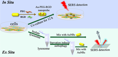
Autophagy plays a critical role in many vitally important physiological and pathological processes, such as the removal of damaged and aged organelles and redundant proteins. Although autophagy is mainly a protective process for cells, it can also cause cell death. In this study, we employed in situ and ex situ surface-enhanced Raman scattering (SERS) spectroscopies to obtain chemical information of lysosomes of HepG2 cells. Results reveal that the SERS profiles of the isolated lysosomes are different from the in situ spectra, indicating that lysosomes lie in different microenvironments in these two cases. We further investigated the molecular changes of isolated lysosomes according to the autophagy induced by starvation viaex situ SERS. During autophagy, the conformation of proteins and the structures of lipids have been affected, and autophagy-related molecular evidence is given for the first time in the living lysosomes. We expect that this study will provide a reference for understanding the cell autophagy mechanism.
中文翻译:

通过表面增强拉曼散射光谱研究溶酶体自噬
自噬在许多至关重要的生理和病理过程中起着至关重要的作用,例如去除受损和老化的细胞器和多余的蛋白质。尽管自噬主要是对细胞的保护过程,但它也会导致细胞死亡。在这项研究中,我们采用原位和异位表面增强拉曼散射 (SERS) 光谱来获取 HepG2 细胞溶酶体的化学信息。结果表明,分离出的溶酶体的 SERS 谱与原位光谱不同,表明在这两种情况下溶酶体位于不同的微环境中。根据由饥饿诱导自噬我们进一步研究分离溶酶体的分子变化经由易地伺服器。自噬过程中,蛋白质的构象和脂质的结构受到影响,首次在活的溶酶体中给出了与自噬相关的分子证据。期待本研究为了解细胞自噬机制提供参考。
更新日期:2021-09-28
中文翻译:

通过表面增强拉曼散射光谱研究溶酶体自噬
自噬在许多至关重要的生理和病理过程中起着至关重要的作用,例如去除受损和老化的细胞器和多余的蛋白质。尽管自噬主要是对细胞的保护过程,但它也会导致细胞死亡。在这项研究中,我们采用原位和异位表面增强拉曼散射 (SERS) 光谱来获取 HepG2 细胞溶酶体的化学信息。结果表明,分离出的溶酶体的 SERS 谱与原位光谱不同,表明在这两种情况下溶酶体位于不同的微环境中。根据由饥饿诱导自噬我们进一步研究分离溶酶体的分子变化经由易地伺服器。自噬过程中,蛋白质的构象和脂质的结构受到影响,首次在活的溶酶体中给出了与自噬相关的分子证据。期待本研究为了解细胞自噬机制提供参考。


 京公网安备 11010802027423号