当前位置:
X-MOL 学术
›
J. Neurosci. Res.
›
论文详情
Our official English website, www.x-mol.net, welcomes your feedback! (Note: you will need to create a separate account there.)
Hippocampal and striatal volumes correlate with spatial memory impairment in Huntington’s disease
Journal of Neuroscience Research ( IF 4.2 ) Pub Date : 2021-09-13 , DOI: 10.1002/jnr.24966 Yifat Glikmann-Johnston 1 , Emily-Clare Mercieca 1 , Anna M Carmichael 1 , Bonnie Alexander 1, 2, 3 , Ian H Harding 4, 5 , Julie C Stout 1
Journal of Neuroscience Research ( IF 4.2 ) Pub Date : 2021-09-13 , DOI: 10.1002/jnr.24966 Yifat Glikmann-Johnston 1 , Emily-Clare Mercieca 1 , Anna M Carmichael 1 , Bonnie Alexander 1, 2, 3 , Ian H Harding 4, 5 , Julie C Stout 1
Affiliation
|
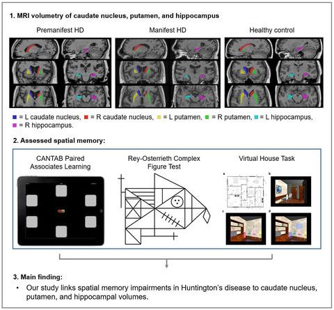
Spatial memory impairments are observed in people with Huntington's disease (HD), however, the domain of spatial memory has received little focus when characterizing the cognitive phenotype of HD. Spatial memory is traditionally thought to be a hippocampal-dependent function, while the neuropathology of HD centers on the striatum. Alongside spatial memory deficits in HD, recent neurocognitive theories suggest that a larger brain network is involved, including the striatum. We examined the relationship between hippocampal and striatal volumes and spatial memory in 36 HD gene expansion carriers, including premanifest (n = 24) and early manifest HD (n = 12), and 32 matched healthy controls. We assessed spatial memory with Paired Associates Learning, Rey–Osterrieth Complex Figure Test, and the Virtual House task, which assesses three components of spatial memory: navigation, object location, and plan drawing. Caudate nucleus, putamen, and hippocampal volumes were manually segmented on T1-weighted MR images. As expected, caudate nucleus and putamen volumes were significantly smaller in the HD group compared to controls, with manifest HD having more severe atrophy than the premanifest HD group. Hippocampal volumes did not differ significantly between HD and control groups. Nonetheless, on average, the HD group performed significantly worse than controls across all spatial memory tasks. The spatial memory components of object location and recall of figural and topographical drawings were associated with striatal and hippocampal volumes in the HD cohort. We provide a case to include spatial memory impairments in the cognitive phenotype of HD, and extend the neurocognitive picture of HD beyond its primary pathology within the striatum.
中文翻译:

海马和纹状体体积与亨廷顿病的空间记忆障碍相关
在亨廷顿病 (HD) 患者中观察到空间记忆障碍,然而,在表征 HD 的认知表型时,空间记忆领域几乎没有受到关注。空间记忆传统上被认为是海马依赖的功能,而 HD 的神经病理学集中在纹状体上。除了 HD 中的空间记忆缺陷外,最近的神经认知理论表明涉及更大的大脑网络,包括纹状体。我们检查了 36 名 HD 基因扩增携带者的海马和纹状体体积与空间记忆之间的关系,包括表现前 ( n = 24) 和早期表现 HD ( n = 12) 和 32 名匹配的健康对照。我们使用 Paired Associates Learning、Rey-Osterrieth Complex Figure Test 和 Virtual House 任务评估了空间记忆,该任务评估了空间记忆的三个组成部分:导航、对象位置和平面图。在 T1 加权 MR 图像上手动分割尾状核、壳核和海马体积。正如预期的那样,与对照组相比,HD 组的尾状核和壳核体积明显更小,明显的 HD 比表现前的 HD 组具有更严重的萎缩。HD 组和对照组之间的海马体积没有显着差异。尽管如此,平均而言,HD 组在所有空间记忆任务中的表现都比对照组差得多。对象位置的空间记忆成分以及对图形和地形图的回忆与 HD 队列中的纹状体和海马体积相关。我们提供了一个案例,将空间记忆障碍包括在 HD 的认知表型中,并将 HD 的神经认知图像扩展到纹状体内的主要病理之外。
更新日期:2021-09-13
中文翻译:
海马和纹状体体积与亨廷顿病的空间记忆障碍相关
在亨廷顿病 (HD) 患者中观察到空间记忆障碍,然而,在表征 HD 的认知表型时,空间记忆领域几乎没有受到关注。空间记忆传统上被认为是海马依赖的功能,而 HD 的神经病理学集中在纹状体上。除了 HD 中的空间记忆缺陷外,最近的神经认知理论表明涉及更大的大脑网络,包括纹状体。我们检查了 36 名 HD 基因扩增携带者的海马和纹状体体积与空间记忆之间的关系,包括表现前 ( n = 24) 和早期表现 HD ( n = 12) 和 32 名匹配的健康对照。我们使用 Paired Associates Learning、Rey-Osterrieth Complex Figure Test 和 Virtual House 任务评估了空间记忆,该任务评估了空间记忆的三个组成部分:导航、对象位置和平面图。在 T1 加权 MR 图像上手动分割尾状核、壳核和海马体积。正如预期的那样,与对照组相比,HD 组的尾状核和壳核体积明显更小,明显的 HD 比表现前的 HD 组具有更严重的萎缩。HD 组和对照组之间的海马体积没有显着差异。尽管如此,平均而言,HD 组在所有空间记忆任务中的表现都比对照组差得多。对象位置的空间记忆成分以及对图形和地形图的回忆与 HD 队列中的纹状体和海马体积相关。我们提供了一个案例,将空间记忆障碍包括在 HD 的认知表型中,并将 HD 的神经认知图像扩展到纹状体内的主要病理之外。


 京公网安备 11010802027423号