Our official English website, www.x-mol.net, welcomes your feedback! (Note: you will need to create a separate account there.)
Review: tomographic imaging flow cytometry
Lab on a Chip ( IF 6.1 ) Pub Date : 2021-09-13 , DOI: 10.1039/d1lc00533b Andreas Kleiber 1 , Daniel Kraus 1 , Thomas Henkel 1 , Wolfgang Fritzsche 1
Lab on a Chip ( IF 6.1 ) Pub Date : 2021-09-13 , DOI: 10.1039/d1lc00533b Andreas Kleiber 1 , Daniel Kraus 1 , Thomas Henkel 1 , Wolfgang Fritzsche 1
Affiliation
|
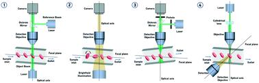
Within the last decades, conventional flow cytometry (FC) has evolved as a powerful measurement method in clinical diagnostics, biology, life sciences and healthcare. Imaging flow cytometry (IFC) extends the power of traditional FC by adding high resolution optical and spectroscopic information. However, the conventional IFC only provides a 2D projection of a 3D object. To overcome this limitation, tomographic imaging flow cytometry (tIFC) was developed to access 3D information about the target particles. The goal of tIFC is to visualize surfaces and internal structures in a holistic way. This review article gives an overview of the past and current developments in tIFC.
中文翻译:

评论:断层成像流式细胞术
在过去的几十年里,传统的流式细胞术 (FC) 已经发展成为临床诊断、生物学、生命科学和医疗保健领域的一种强大的测量方法。成像流式细胞术 (IFC) 通过添加高分辨率光学和光谱信息扩展了传统 FC 的功能。然而,传统的 IFC 仅提供 3D 对象的 2D 投影。为了克服这一限制,开发了断层成像流式细胞术 (tIFC) 来获取有关目标粒子的 3D 信息。tIFC 的目标是以整体方式可视化表面和内部结构。这篇评论文章概述了 tIFC 的过去和当前发展。
更新日期:2021-09-13
中文翻译:
评论:断层成像流式细胞术
在过去的几十年里,传统的流式细胞术 (FC) 已经发展成为临床诊断、生物学、生命科学和医疗保健领域的一种强大的测量方法。成像流式细胞术 (IFC) 通过添加高分辨率光学和光谱信息扩展了传统 FC 的功能。然而,传统的 IFC 仅提供 3D 对象的 2D 投影。为了克服这一限制,开发了断层成像流式细胞术 (tIFC) 来获取有关目标粒子的 3D 信息。tIFC 的目标是以整体方式可视化表面和内部结构。这篇评论文章概述了 tIFC 的过去和当前发展。


 京公网安备 11010802027423号