Our official English website, www.x-mol.net, welcomes your feedback! (Note: you will need to create a separate account there.)
Terpyridine Zn(II) Complexes with Azide Units for Visualization of Histone Deacetylation in Living Cells under STED Nanoscopy
ACS Sensors ( IF 8.9 ) Pub Date : 2021-09-09 , DOI: 10.1021/acssensors.1c01287 Wei Du 1, 2 , Dayi Pan 1 , Pan Xiang 1 , Chaoya Xiong 3 , Mingzhu Zhang 3 , Qiong Zhang 3 , Yupeng Tian 3 , Zhongping Zhang 3, 4 , Bo Chen 5 , Kui Luo 1, 6 , Qiyong Gong 1, 6 , Xiaohe Tian 1, 3, 6
ACS Sensors ( IF 8.9 ) Pub Date : 2021-09-09 , DOI: 10.1021/acssensors.1c01287 Wei Du 1, 2 , Dayi Pan 1 , Pan Xiang 1 , Chaoya Xiong 3 , Mingzhu Zhang 3 , Qiong Zhang 3 , Yupeng Tian 3 , Zhongping Zhang 3, 4 , Bo Chen 5 , Kui Luo 1, 6 , Qiyong Gong 1, 6 , Xiaohe Tian 1, 3, 6
Affiliation

|
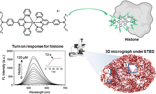
Histones are the alkali proteins in eukaryotic somatic chromatin cells which constitute the nucleosome structure together with DNA. Their abnormality is often associated with multiple tumorigenesis and other human diseases. Nevertheless, a simple and efficient super-resolution method to visualize histone distribution at the subcellular level is still unavailable. Herein, a Zn(II) terpyridine complex with rich-electronic azide units, namely, TpZnA–His, was designed and synthesized. The initial in vitro and in silico studies suggested that this complex is able to detect histones rapidly and selectively via charge–charge interactions with the histone H3 subunit. Its live cell nuclear localization, red-emission tail, and large Stokes shift allowed super-resolution evaluation of histone distributions with a clear distinction against nuclear DNA. We were able to quantitatively conclude three histone morphology alternations in live cells including condensation, aggregation, and cavity during activating histone acetylation. This work offers a better understanding as well as a versatile tool to study histone-involved gene transcription, signal transduction, and differentiation in cells.
中文翻译:

具有叠氮单元的三联吡啶锌 (II) 配合物用于在 STED 纳米镜下观察活细胞中的组蛋白去乙酰化
组蛋白是真核体染色质细胞中的碱性蛋白质,与 DNA 一起构成核小体结构。它们的异常通常与多种肿瘤发生和其他人类疾病有关。然而,在亚细胞水平上可视化组蛋白分布的简单有效的超分辨率方法仍然不可用。在此,设计并合成了一种具有丰富电子叠氮化物单元的 Zn(II) 三联吡啶配合物,即 TpZnA-His。最初的体外和计算机研究表明,这种复合物能够通过以下方式快速和选择性地检测组蛋白与组蛋白 H3 亚基的电荷-电荷相互作用。其活细胞核定位、红光尾和大斯托克斯位移允许对组蛋白分布进行超分辨率评估,并与核 DNA 有明显区别。我们能够定量总结活细胞中的三种组蛋白形态变化,包括在激活组蛋白乙酰化过程中的凝聚、聚集和空洞。这项工作提供了更好的理解以及研究细胞中组蛋白相关基因转录、信号转导和分化的通用工具。
更新日期:2021-09-09
中文翻译:

具有叠氮单元的三联吡啶锌 (II) 配合物用于在 STED 纳米镜下观察活细胞中的组蛋白去乙酰化
组蛋白是真核体染色质细胞中的碱性蛋白质,与 DNA 一起构成核小体结构。它们的异常通常与多种肿瘤发生和其他人类疾病有关。然而,在亚细胞水平上可视化组蛋白分布的简单有效的超分辨率方法仍然不可用。在此,设计并合成了一种具有丰富电子叠氮化物单元的 Zn(II) 三联吡啶配合物,即 TpZnA-His。最初的体外和计算机研究表明,这种复合物能够通过以下方式快速和选择性地检测组蛋白与组蛋白 H3 亚基的电荷-电荷相互作用。其活细胞核定位、红光尾和大斯托克斯位移允许对组蛋白分布进行超分辨率评估,并与核 DNA 有明显区别。我们能够定量总结活细胞中的三种组蛋白形态变化,包括在激活组蛋白乙酰化过程中的凝聚、聚集和空洞。这项工作提供了更好的理解以及研究细胞中组蛋白相关基因转录、信号转导和分化的通用工具。


 京公网安备 11010802027423号