Journal of the American Society of Echocardiography ( IF 6.5 ) Pub Date : 2021-09-08 , DOI: 10.1016/j.echo.2021.08.023 Pedro Morais 1 , Yiting Fan 2 , Sandro Queirós 3 , Jan D'hooge 4 , Alex Pui-Wai Lee 5 , João L Vilaça 1
|
Background
Procedural success of transcatheter left atrial appendage closure (LAAC) is dependent on correct device selection. Three-dimensional (3D) transesophageal echocardiography (TEE) is more accurate than the two-dimensional modality for evaluation of the complex anatomy of the left atrial appendage (LAA). However, 3D transesophageal echocardiographic analysis of the LAA is challenging and highly expertise dependent. The aim of this study was to evaluate the feasibility and accuracy of a novel software tool for automated 3D analysis of the LAA using 3D transesophageal echocardiographic data.
Methods
Intraprocedural 3D transesophageal echocardiographic data from 158 patients who underwent LAAC were retrospectively analyzed using a novel automated LAA analysis software tool. On the basis of the 3D transesophageal echocardiographic data, the software semiautomatically segmented the 3D LAA structure, determined the device landing zone, and generated measurements of the landing zone dimensions and LAA length, allowing manual editing if necessary. The accuracy of LAA preimplantation anatomic measurement reproducibility and time for analysis of the automated software were compared against expert manual 3D analysis. The software feasibility to predict the optimal device size was directly compared with implanted models.
Results
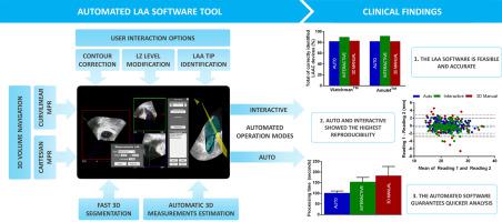
Automated 3D analysis of the LAA on 3D TEE was feasible in all patients. There was excellent agreement between automated and manual measurements of landing zone maximal diameter (bias, −0.32; limits of agreement, −3.56 to 2.92), area-derived mean diameter (bias, −0.24; limits of agreement, −3.12 to 2.64), and LAA depth (bias, 0.02; limits of agreement, −3.14 to 3.18). Automated 3D analysis, with manual editing if necessary, accurately identified the implanted device size in 90.5% of patients, outperforming two-dimensional TEE (68.9%; P < .01). The automated software showed results competitive against the manual analysis of 3D TEE, with higher intra- and interobserver reproducibility, and allowed quicker analysis (101.9 ± 9.3 vs 183.5 ± 42.7 sec, P < .001) compared with manual analysis.
Conclusions
Automated LAA analysis on the basis of 3D TEE is feasible and allows accurate, reproducible, and rapid device sizing decision for LAAC.
中文翻译:
经导管封堵左心耳自动三维超声心动图分析的可行性和准确性
背景
经导管左心耳封堵术 (LAAC) 的手术成功取决于正确的器械选择。三维 (3D) 经食道超声心动图 (TEE) 在评估左心耳 (LAA) 的复杂解剖结构时比二维模式更准确。然而,LAA 的 3D 经食道超声心动图分析具有挑战性并且高度依赖专业知识。本研究的目的是评估使用 3D 经食道超声心动图数据对 LAA 进行自动 3D 分析的新型软件工具的可行性和准确性。
方法
使用新型自动 LAA 分析软件工具对 158 名接受 LAAC 的患者的术中 3D 经食道超声心动图数据进行回顾性分析。在 3D 经食道超声心动图数据的基础上,软件半自动分割 3D LAA 结构,确定设备着陆区,并生成着陆区尺寸和 LAA 长度的测量值,必要时允许手动编辑。LAA 植入前解剖测量的准确性和自动化软件分析的时间与专家手动 3D 分析进行了比较。预测最佳设备尺寸的软件可行性直接与植入模型进行比较。
结果
在 3D TEE 上对 LAA 进行自动 3D 分析在所有患者中都是可行的。着陆区最大直径(偏差,-0.32;一致性极限,-3.56 到 2.92)、面积衍生的平均直径(偏差,-0.24;一致性极限,-3.12 到 2.64)的自动和手动测量结果非常一致和 LAA 深度(偏差,0.02;一致性限制,-3.14 到 3.18)。自动化 3D 分析,必要时进行手动编辑,准确识别了 90.5% 患者的植入装置尺寸,优于二维 TEE (68.9%;P < .01)。自动化软件显示出与 3D TEE 手动分析相比具有竞争力的结果,具有更高的观察者内部和观察者间重现性,并允许更快的分析(101.9 ± 9.3 对 183.5 ± 42.7 秒,P < .001) 与手动分析相比。
结论
基于 3D TEE 的自动 LAA 分析是可行的,并且可以为 LAAC 做出准确、可重复和快速的设备尺寸决策。


 京公网安备 11010802027423号