当前位置:
X-MOL 学术
›
ACS Appl. Bio Mater.
›
论文详情
Our official English website, www.x-mol.net, welcomes your feedback! (Note: you will need to create a separate account there.)
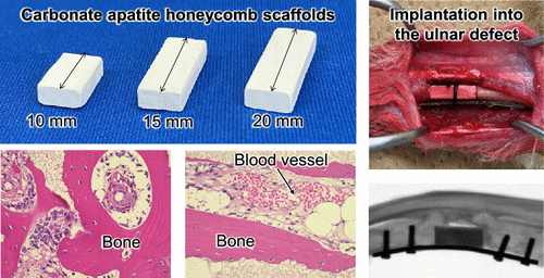
Honeycomb Scaffold-Guided Bone Reconstruction of Critical-Sized Defects in Rabbit Ulnar Shafts
ACS Applied Bio Materials ( IF 4.7 ) Pub Date : 2021-09-02 , DOI: 10.1021/acsabm.1c00533 Keigo Shibahara 1, 2 , Koichiro Hayashi 2 , Yasuharu Nakashima 1 , Kunio Ishikawa 2
ACS Applied Bio Materials ( IF 4.7 ) Pub Date : 2021-09-02 , DOI: 10.1021/acsabm.1c00533 Keigo Shibahara 1, 2 , Koichiro Hayashi 2 , Yasuharu Nakashima 1 , Kunio Ishikawa 2
Affiliation
|
Reconstruction of critical-sized defects (CSDs) in bone shafts remains a major challenge in orthopedics. Honeycomb (HC) scaffolds are considered promising as their uniaxial channels bridge the amputation stumps of bones and promote the ingrowth of bone and blood vessels (BV) into the scaffolds. In this study, the ability of the HC scaffolds, composed of the bone mineral or carbonate apatite (CAp), was evaluated by reconstructing 10, 15, and 20 mm segmental defects in the rabbit ulnar shaft. Radiographic and μ-computed tomography evaluations showed that bony calluses were formed around the scaffolds at 4 weeks post-surgery in all defects, whereas no callus bridged in the ulna without scaffolds. At 12 weeks post-surgery, the scaffolds were connected to the host bone in 10 and 15 mm defects, while a slight gap remained between the scaffold and host bone in the 20 mm defect. New bone formation and scaffold resorption progressed over 12 weeks. Histological evaluations showed that mature bones (MB) and BV were already formed at the edges of the scaffolds at 4 weeks post-surgery in 10, 15, and 20 mm defects. In the central region of the scaffold, in the 10 mm defect, MB and BV were formed at 4 weeks post-surgery. In the 15 mm defect, although BV were formed, a few MB were formed. It is concluded that CAp HC scaffolds have good potential value for the reconstruction of CSDs.
中文翻译:

蜂窝支架引导骨重建兔尺骨临界尺寸缺损
骨干中临界尺寸缺陷 (CSD) 的重建仍然是骨科面临的主要挑战。蜂窝 (HC) 支架被认为是有前途的,因为它们的单轴通道桥接骨骼的截肢残端并促进骨骼和血管 (BV) 向支架内生长。在这项研究中,通过重建兔尺骨干中 10、15 和 20 mm 的节段性缺损来评估由骨矿物质或碳酸盐磷灰石 (CAp) 组成的 HC 支架的能力。射线照相和 μ 计算机断层扫描评估显示,在所有缺损处术后 4 周,支架周围均形成骨性愈伤组织,而没有支架的尺骨中没有愈伤组织桥接。术后 12 周,支架在 10 和 15 毫米缺损处与宿主骨连接,而在 20 mm 缺损处,支架和宿主骨之间仍存在微小间隙。新骨形成和支架吸收进展超过 12 周。组织学评估表明,在 10、15 和 20 毫米缺损处,手术后 4 周,支架边缘已经形成成熟骨 (MB) 和 BV。在支架的中心区域,在 10 mm 缺损处,MB 和 BV 在术后 4 周形成。在 15 mm 的缺损处,虽然形成了 BV,但也形成了一些 MB。得出结论,CAP HC支架对于CSD的重建具有良好的潜在价值。在支架的中心区域,在 10 mm 缺损处,MB 和 BV 在术后 4 周形成。在 15 mm 的缺损处,虽然形成了 BV,但也形成了一些 MB。得出结论,CAP HC支架对于CSD的重建具有良好的潜在价值。在支架的中心区域,在 10 mm 缺损处,MB 和 BV 在术后 4 周形成。在 15 mm 的缺损处,虽然形成了 BV,但也形成了一些 MB。得出结论,CAP HC支架对于CSD的重建具有良好的潜在价值。
更新日期:2021-09-20
中文翻译:
蜂窝支架引导骨重建兔尺骨临界尺寸缺损
骨干中临界尺寸缺陷 (CSD) 的重建仍然是骨科面临的主要挑战。蜂窝 (HC) 支架被认为是有前途的,因为它们的单轴通道桥接骨骼的截肢残端并促进骨骼和血管 (BV) 向支架内生长。在这项研究中,通过重建兔尺骨干中 10、15 和 20 mm 的节段性缺损来评估由骨矿物质或碳酸盐磷灰石 (CAp) 组成的 HC 支架的能力。射线照相和 μ 计算机断层扫描评估显示,在所有缺损处术后 4 周,支架周围均形成骨性愈伤组织,而没有支架的尺骨中没有愈伤组织桥接。术后 12 周,支架在 10 和 15 毫米缺损处与宿主骨连接,而在 20 mm 缺损处,支架和宿主骨之间仍存在微小间隙。新骨形成和支架吸收进展超过 12 周。组织学评估表明,在 10、15 和 20 毫米缺损处,手术后 4 周,支架边缘已经形成成熟骨 (MB) 和 BV。在支架的中心区域,在 10 mm 缺损处,MB 和 BV 在术后 4 周形成。在 15 mm 的缺损处,虽然形成了 BV,但也形成了一些 MB。得出结论,CAP HC支架对于CSD的重建具有良好的潜在价值。在支架的中心区域,在 10 mm 缺损处,MB 和 BV 在术后 4 周形成。在 15 mm 的缺损处,虽然形成了 BV,但也形成了一些 MB。得出结论,CAP HC支架对于CSD的重建具有良好的潜在价值。在支架的中心区域,在 10 mm 缺损处,MB 和 BV 在术后 4 周形成。在 15 mm 的缺损处,虽然形成了 BV,但也形成了一些 MB。得出结论,CAP HC支架对于CSD的重建具有良好的潜在价值。


 京公网安备 11010802027423号