当前位置:
X-MOL 学术
›
J. Neurochem.
›
论文详情
Our official English website, www.x-mol.net, welcomes your feedback! (Note: you will need to create a separate account there.)
Spontaneous, transient adenosine release is not enhanced in the CA1 region of hippocampus during severe ischemia models
Journal of Neurochemistry ( IF 4.7 ) Pub Date : 2021-08-28 , DOI: 10.1111/jnc.15496 Mallikarjunarao Ganesana 1 , B Jill Venton 1
Journal of Neurochemistry ( IF 4.7 ) Pub Date : 2021-08-28 , DOI: 10.1111/jnc.15496 Mallikarjunarao Ganesana 1 , B Jill Venton 1
Affiliation
|
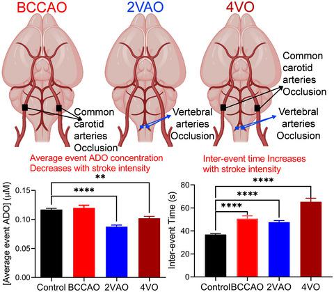
Ischemic stroke causes damage in the brain, and a slow buildup of adenosine is neuroprotective during ischemic injury. Spontaneous, transient adenosine signaling, lasting only 3 s per event, has been discovered that increases in frequency in the caudate-putamen during early stages of mild ischemia-reperfusion injury. However, spontaneous adenosine changes have not been studied in the hippocampus during ischemia, an area highly susceptible to stroke. Here, we investigated changes of spontaneous, transient adenosine in the CA1 region of rat hippocampus during three different models of the varied intensity of ischemia. During the early stages of the milder bilateral common carotid artery occlusion (BCCAO) model, there were fewer spontaneous, transient adenosine, but no change in the concentration of individual events. In contrast, during the moderate 2 vertebral artery occlusion (2VAO) and severe 4 vessel occlusion (4VO) models, both the frequency of spontaneous, transient adenosine and the average event adenosine concentration decreased. Blood flow measurements validate that the ischemia models decreased blood flow, and corresponding pathological changes were observed by transmission electron microscopy (TEM). 4VO occlusion showed the most severe damage in histology and BCCAO showed the least. Overall, our data suggest that there is no enhanced spontaneous adenosine release in the hippocampus during moderate and severe ischemia, which could be due to depletion of the rapidly releasable adenosine pool. Thus, during ischemic stroke, there are fewer spontaneous adenosine events that could inhibit neurotransmission, which might lead to more damage and less neuroprotection in the hippocampus CA1 region.
中文翻译:

在严重缺血模型中,海马 CA1 区的自发性瞬时腺苷释放没有增强
缺血性中风会导致大脑受损,而腺苷的缓慢积累在缺血性损伤期间具有神经保护作用。自发的、短暂的腺苷信号,每次事件仅持续 3 秒,已被发现在轻度缺血再灌注损伤的早期阶段尾壳核的频率增加。然而,尚未研究缺血期间海马体中自发的腺苷变化,海马体是一个极易发生中风的区域。在这里,我们研究了在不同强度缺血的三种不同模型中大鼠海马 CA1 区自发性瞬时腺苷的变化。在较轻的双侧颈总动脉闭塞 (BCCAO) 模型的早期阶段,自发性瞬时腺苷较少,但个别事件的浓度没有变化。相比之下,在中度 2 椎动脉闭塞 (2VAO) 和重度 4 血管闭塞 (4VO) 模型中,自发性、瞬时腺苷的频率和平均事件腺苷浓度均降低。血流测量证实了缺血模型降低了血流,并通过透射电子显微镜(TEM)观察到相应的病理变化。4VO闭塞组织学损伤最严重,BCCAO最轻。总体而言,我们的数据表明,在中度和重度缺血期间,海马体中的自发腺苷释放没有增强,这可能是由于可快速释放的腺苷池耗尽所致。因此,在缺血性中风期间,可抑制神经传递的自发性腺苷事件较少,
更新日期:2021-08-28
中文翻译:
在严重缺血模型中,海马 CA1 区的自发性瞬时腺苷释放没有增强
缺血性中风会导致大脑受损,而腺苷的缓慢积累在缺血性损伤期间具有神经保护作用。自发的、短暂的腺苷信号,每次事件仅持续 3 秒,已被发现在轻度缺血再灌注损伤的早期阶段尾壳核的频率增加。然而,尚未研究缺血期间海马体中自发的腺苷变化,海马体是一个极易发生中风的区域。在这里,我们研究了在不同强度缺血的三种不同模型中大鼠海马 CA1 区自发性瞬时腺苷的变化。在较轻的双侧颈总动脉闭塞 (BCCAO) 模型的早期阶段,自发性瞬时腺苷较少,但个别事件的浓度没有变化。相比之下,在中度 2 椎动脉闭塞 (2VAO) 和重度 4 血管闭塞 (4VO) 模型中,自发性、瞬时腺苷的频率和平均事件腺苷浓度均降低。血流测量证实了缺血模型降低了血流,并通过透射电子显微镜(TEM)观察到相应的病理变化。4VO闭塞组织学损伤最严重,BCCAO最轻。总体而言,我们的数据表明,在中度和重度缺血期间,海马体中的自发腺苷释放没有增强,这可能是由于可快速释放的腺苷池耗尽所致。因此,在缺血性中风期间,可抑制神经传递的自发性腺苷事件较少,


 京公网安备 11010802027423号