当前位置:
X-MOL 学术
›
J. Leukoc. Biol.
›
论文详情
Our official English website, www.x-mol.net, welcomes your feedback! (Note: you will need to create a separate account there.)
Pulmonary and blood dendritic cells from sarcoidosis patients more potently induce IFNγ-producing Th1 cells compared with monocytes
Journal of Leukocyte Biology ( IF 5.5 ) Pub Date : 2021-08-25 , DOI: 10.1002/jlb.5a0321-162r Rico Lepzien 1 , Mu Nie 1 , Paulo Czarnewski 2 , Sang Liu 1 , Meng Yu 1 , Avinash Ravindran 3 , Susanna Kullberg 3, 4 , Anders Eklund 3, 4 , Johan Grunewald 3, 4 , Anna Smed-Sörensen 1
Journal of Leukocyte Biology ( IF 5.5 ) Pub Date : 2021-08-25 , DOI: 10.1002/jlb.5a0321-162r Rico Lepzien 1 , Mu Nie 1 , Paulo Czarnewski 2 , Sang Liu 1 , Meng Yu 1 , Avinash Ravindran 3 , Susanna Kullberg 3, 4 , Anders Eklund 3, 4 , Johan Grunewald 3, 4 , Anna Smed-Sörensen 1
Affiliation
|
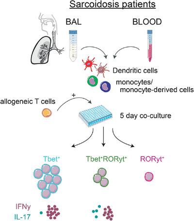
Sarcoidosis is a systemic inflammatory disease mainly affecting the lungs. The hallmark of sarcoidosis are granulomas that are surrounded by activated T cells, likely targeting the disease-inducing antigen. IFNγ-producing Th1 and Th17.1 T cells are elevated in sarcoidosis and associate with disease progression. Monocytes and dendritic cells (DCs) are antigen-presenting cells (APCs) and required for T cell activation. Several subsets of monocytes and DCs with different functions were identified in sarcoidosis. However, to what extent different monocyte and DC subsets can support activation and skewing of T cells in sarcoidosis is still unclear. In this study, we performed a transcriptional and functional side-by-side comparison of sorted monocytes and DCs from matched blood and bronchoalveolar lavage (BAL) fluid of sarcoidosis patients. Transcriptomic analysis of all subsets showed upregulation of genes related to T cell activation and antigen presentation in DCs compared with monocytes. Allogeneic T cell proliferation was higher after coculture with monocytes and DCs from blood compared with BAL and DCs induced more T cell proliferation compared with monocytes. After coculture, proliferating T cells showed high expression of the transcription factor Tbet and IFNγ production. We also identified Tbet and RORγt coexpressing T cells that mainly produced IFNγ. Our data show that DCs rather than monocytes from sarcoidosis patients have the ability to activate and polarize T cells towards Th1 and Th17.1 cells. This study provides a useful in vitro tool to better understand the contribution of monocytes and DCs to T cell activation and immunopathology in sarcoidosis.
中文翻译:

与单核细胞相比,来自结节病患者的肺和血液树突状细胞更有效地诱导产生 IFNγ 的 Th1 细胞
结节病是一种全身性炎症性疾病,主要影响肺部。结节病的标志是被活化的 T 细胞包围的肉芽肿,很可能靶向诱导疾病的抗原。产生 IFNγ 的 Th1 和 Th17.1 T 细胞在结节病中升高并与疾病进展相关。单核细胞和树突状细胞 (DC) 是抗原呈递细胞 (APC),是 T 细胞活化所必需的。在结节病中鉴定出几个具有不同功能的单核细胞和 DCs 亚群。然而,在何种程度上不同的单核细胞和 DC 亚群可以支持结节病中 T 细胞的激活和偏斜仍不清楚。在这项研究中,我们对来自结节病患者的匹配血液和支气管肺泡灌洗液 (BAL) 液的分选单核细胞和 DCs 进行了转录和功能并排比较。所有亚群的转录组学分析显示,与单核细胞相比,DC 中与 T 细胞活化和抗原呈递相关的基因上调。与 BAL 相比,与来自血液的单核细胞和 DCs 共培养后同种异体 T 细胞增殖更高,与单核细胞相比,DCs 诱导更多的 T 细胞增殖。共培养后,增殖的 T 细胞表现出转录因子 Tbet 的高表达和 IFNγ 的产生。我们还鉴定了主要产生 IFNγ 的 Tbet 和 RORγt 共表达 T 细胞。我们的数据表明,来自结节病患者的 DC 而不是单核细胞具有激活 T 细胞并将其极化为 Th1 和 Th17.1 细胞的能力。本研究提供了一种有用的体外工具,可以更好地了解单核细胞和 DCs 对结节病中 T 细胞活化和免疫病理学的贡献。
更新日期:2021-08-25
中文翻译:
与单核细胞相比,来自结节病患者的肺和血液树突状细胞更有效地诱导产生 IFNγ 的 Th1 细胞
结节病是一种全身性炎症性疾病,主要影响肺部。结节病的标志是被活化的 T 细胞包围的肉芽肿,很可能靶向诱导疾病的抗原。产生 IFNγ 的 Th1 和 Th17.1 T 细胞在结节病中升高并与疾病进展相关。单核细胞和树突状细胞 (DC) 是抗原呈递细胞 (APC),是 T 细胞活化所必需的。在结节病中鉴定出几个具有不同功能的单核细胞和 DCs 亚群。然而,在何种程度上不同的单核细胞和 DC 亚群可以支持结节病中 T 细胞的激活和偏斜仍不清楚。在这项研究中,我们对来自结节病患者的匹配血液和支气管肺泡灌洗液 (BAL) 液的分选单核细胞和 DCs 进行了转录和功能并排比较。所有亚群的转录组学分析显示,与单核细胞相比,DC 中与 T 细胞活化和抗原呈递相关的基因上调。与 BAL 相比,与来自血液的单核细胞和 DCs 共培养后同种异体 T 细胞增殖更高,与单核细胞相比,DCs 诱导更多的 T 细胞增殖。共培养后,增殖的 T 细胞表现出转录因子 Tbet 的高表达和 IFNγ 的产生。我们还鉴定了主要产生 IFNγ 的 Tbet 和 RORγt 共表达 T 细胞。我们的数据表明,来自结节病患者的 DC 而不是单核细胞具有激活 T 细胞并将其极化为 Th1 和 Th17.1 细胞的能力。本研究提供了一种有用的体外工具,可以更好地了解单核细胞和 DCs 对结节病中 T 细胞活化和免疫病理学的贡献。


 京公网安备 11010802027423号