当前位置:
X-MOL 学术
›
Immunology
›
论文详情
Our official English website, www.x-mol.net, welcomes your feedback! (Note: you will need to create a separate account there.)
Memory CD8+ T cells exhibit tissue imprinting and non-stable exposure-dependent reactivation characteristics following blood-stage Plasmodium berghei ANKA infections
Immunology ( IF 6.4 ) Pub Date : 2021-08-18 , DOI: 10.1111/imm.13405 Tovah N Shaw 1, 2 , Michael J Haley 1 , Rebecca S Dookie 1 , Jenna J Godfrey 1 , Antonn J Cheeseman 1 , Patrick Strangward 1 , Leo A H Zeef 3 , Ana Villegas-Mendez 1 , Kevin N Couper 1
Immunology ( IF 6.4 ) Pub Date : 2021-08-18 , DOI: 10.1111/imm.13405 Tovah N Shaw 1, 2 , Michael J Haley 1 , Rebecca S Dookie 1 , Jenna J Godfrey 1 , Antonn J Cheeseman 1 , Patrick Strangward 1 , Leo A H Zeef 3 , Ana Villegas-Mendez 1 , Kevin N Couper 1
Affiliation
|
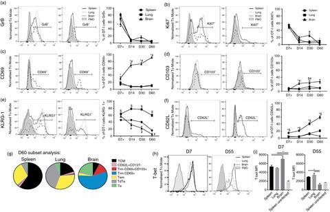
Experimental cerebral malaria (ECM) is a severe complication of Plasmodium berghei ANKA (PbA) infection in mice, characterized by CD8+ T-cell accumulation within the brain. Whilst the dynamics of CD8+ T-cell activation and migration during extant primary PbA infection have been extensively researched, the fate of the parasite-specific CD8+ T cells upon resolution of ECM is not understood. In this study, we show that memory OT-I cells persist systemically within the spleen, lung and brain following recovery from ECM after primary PbA-OVA infection. Whereas memory OT-I cells within the spleen and lung exhibited canonical central memory (Tcm) and effector memory (Tem) phenotypes, respectively, memory OT-I cells within the brain post-PbA-OVA infection displayed an enriched CD69+CD103− profile and expressed low levels of T-bet. OT-I cells within the brain were excluded from short-term intravascular antibody labelling but were targeted effectively by longer-term systemically administered antibodies. Thus, the memory OT-I cells were extravascular within the brain post-ECM but were potentially not resident memory cells. Importantly, whilst memory OT-I cells exhibited strong reactivation during secondary PbA-OVA infection, preventing activation of new primary effector T cells, they had dampened reactivation during a fourth PbA-OVA infection. Overall, our results demonstrate that memory CD8+ T cells are systemically distributed but exhibit a unique phenotype within the brain post-ECM, and that their reactivation characteristics are shaped by infection history. Our results raise important questions regarding the role of distinct memory CD8+ T-cell populations within the brain and other tissues during repeat Plasmodium infections.
中文翻译:

血液阶段伯氏疟原虫 ANKA 感染后,记忆 CD8+ T 细胞表现出组织印记和不稳定的暴露依赖性再激活特征
实验性脑疟疾 (ECM) 是小鼠伯氏疟原虫ANKA (PbA) 感染的严重并发症,其特征在于大脑内的CD8 + T 细胞积聚。虽然在现存的原发性 PbA 感染过程中 CD8 + T 细胞活化和迁移的动态已经被广泛研究,但寄生虫特异性 CD8 +不了解 ECM 分辨率后的 T 细胞。在这项研究中,我们表明记忆 OT-I 细胞在原发性 PbA-OVA 感染后从 ECM 恢复后系统性地存在于脾、肺和脑中。虽然脾脏和肺内的记忆 OT-I 细胞分别表现出典型的中央记忆 (Tcm) 和效应记忆 (Tem) 表型,但脑内 PbA-OVA 感染后的记忆 OT-I 细胞显示出丰富的 CD69 + CD103 -个人资料并表达了低水平的 T-bet。大脑内的 OT-I 细胞被排除在短期血管内抗体标记之外,但被长期全身给药的抗体有效靶向。因此,记忆 OT-I 细胞在 ECM 后脑内血管外,但可能不是常驻记忆细胞。重要的是,虽然记忆 OT-I 细胞在二次 PbA-OVA 感染期间表现出强烈的再激活,阻止了新的初级效应 T 细胞的激活,但它们在第四次 PbA-OVA 感染期间抑制了再激活。总的来说,我们的结果表明内存 CD8 +T 细胞是全身分布的,但在 ECM 后的大脑中表现出独特的表型,并且它们的再激活特征受感染史的影响。我们的结果提出了关于在重复疟原虫感染期间大脑和其他组织内不同记忆 CD8 + T 细胞群的作用的重要问题。
更新日期:2021-08-18
中文翻译:
血液阶段伯氏疟原虫 ANKA 感染后,记忆 CD8+ T 细胞表现出组织印记和不稳定的暴露依赖性再激活特征
实验性脑疟疾 (ECM) 是小鼠伯氏疟原虫ANKA (PbA) 感染的严重并发症,其特征在于大脑内的CD8 + T 细胞积聚。虽然在现存的原发性 PbA 感染过程中 CD8 + T 细胞活化和迁移的动态已经被广泛研究,但寄生虫特异性 CD8 +不了解 ECM 分辨率后的 T 细胞。在这项研究中,我们表明记忆 OT-I 细胞在原发性 PbA-OVA 感染后从 ECM 恢复后系统性地存在于脾、肺和脑中。虽然脾脏和肺内的记忆 OT-I 细胞分别表现出典型的中央记忆 (Tcm) 和效应记忆 (Tem) 表型,但脑内 PbA-OVA 感染后的记忆 OT-I 细胞显示出丰富的 CD69 + CD103 -个人资料并表达了低水平的 T-bet。大脑内的 OT-I 细胞被排除在短期血管内抗体标记之外,但被长期全身给药的抗体有效靶向。因此,记忆 OT-I 细胞在 ECM 后脑内血管外,但可能不是常驻记忆细胞。重要的是,虽然记忆 OT-I 细胞在二次 PbA-OVA 感染期间表现出强烈的再激活,阻止了新的初级效应 T 细胞的激活,但它们在第四次 PbA-OVA 感染期间抑制了再激活。总的来说,我们的结果表明内存 CD8 +T 细胞是全身分布的,但在 ECM 后的大脑中表现出独特的表型,并且它们的再激活特征受感染史的影响。我们的结果提出了关于在重复疟原虫感染期间大脑和其他组织内不同记忆 CD8 + T 细胞群的作用的重要问题。


 京公网安备 11010802027423号