Biomaterials Advances ( IF 7.9 ) Pub Date : 2021-08-19 , DOI: 10.1016/j.msec.2021.112382 Changqi Luo 1 , Claire Wang 2 , Xiangdong Wu 3 , Xiaoping Xie 4 , Chao Wang 5 , Chen Zhao 6 , Chang Zou 7 , Furong Lv 8 , Wei Huang 6 , Junyi Liao 6
|
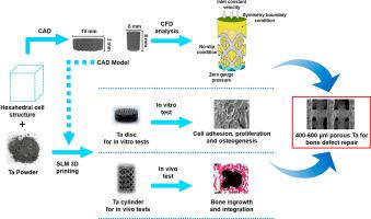
The emerging role of porous tantalum (Ta) scaffold for bone tissue engineering is noticed due to its outstanding biological properties. However, it is controversial which pore size and porosity are more conducive for bone defect repair. In the present work, porous tantalum scaffolds with pore sizes of 100–200, 200–400, 400–600 and 600–800 μm and corresponding porosities of 25%, 55%, 75%, and 85% were constructed, using computer aided design and 3D printing technologies, then comprehensively studied by in vitro and in vivo studies. We found that Ta scaffold with pore size of 400–600 μm showed stronger ability in facilitating cell adhesion, proliferation, and osteogenic differentiation in vitro. In vivo tests identified that porous tantalum scaffolds with pore size of 400–600 μm showed better performance of bone ingrowth and integration. In mechanism, computational fluid dynamics analysis proved porous tantalum scaffolds with pore size of 400–600 μm hold appropriate permeability and surface area, which facilitated cell adhesion and proliferation. Our results strongly indicate that pore size and porosity are essential for further applications of porous tantalum scaffolds, and porous tantalum scaffolds with pore size 400–600 μm are conducive to osteogenesis and osseointegration. These findings provide new evidence for further application of porous tantalum scaffolds for bone defect repair.
中文翻译:
多孔钽支架孔径对成骨和骨整合的影响:基于3D打印技术的综合研究
多孔钽 (Ta) 支架在骨组织工程中的新兴作用因其出色的生物学特性而受到关注。然而,哪种孔径和孔隙率更有利于骨缺损修复存在争议。在目前的工作中,使用计算机辅助构建了孔径为 100-200、200-400、400-600 和 600-800 μm 以及相应孔隙率为 25%、55%、75% 和 85% 的多孔钽支架。设计和 3D 打印技术,然后通过体外和体内研究进行综合研究。我们发现孔径为 400-600 μm 的 Ta 支架在体外促进细胞粘附、增殖和成骨分化的能力更强。体内试验表明,孔径为 400-600 μm 的多孔钽支架显示出更好的骨向内生长和整合性能。在机理上,计算流体动力学分析证明孔径为 400-600 μm 的多孔钽支架具有适当的渗透性和表面积,有利于细胞粘附和增殖。我们的结果强烈表明孔径和孔隙率对于多孔钽支架的进一步应用至关重要,孔径为 400-600 μm 的多孔钽支架有利于成骨和骨整合。这些发现为进一步应用多孔钽支架修复骨缺损提供了新的证据。我们的结果强烈表明孔径和孔隙率对于多孔钽支架的进一步应用至关重要,孔径为 400-600 μm 的多孔钽支架有利于成骨和骨整合。这些发现为进一步应用多孔钽支架修复骨缺损提供了新的证据。我们的结果强烈表明孔径和孔隙率对于多孔钽支架的进一步应用至关重要,孔径为 400-600 μm 的多孔钽支架有利于成骨和骨整合。这些发现为进一步应用多孔钽支架修复骨缺损提供了新的证据。


 京公网安备 11010802027423号