当前位置:
X-MOL 学术
›
Phys. Rev. X
›
论文详情
Our official English website, www.x-mol.net, welcomes your feedback! (Note: you will need to create a separate account there.)
3D Shape of Epithelial Cells on Curved Substrates
Physical Review X ( IF 12.5 ) Pub Date : 2021-08-04 , DOI: 10.1103/physrevx.11.031028 Nicolas Harmand , Anqi Huang , Sylvie Hénon
Physical Review X ( IF 12.5 ) Pub Date : 2021-08-04 , DOI: 10.1103/physrevx.11.031028 Nicolas Harmand , Anqi Huang , Sylvie Hénon
|
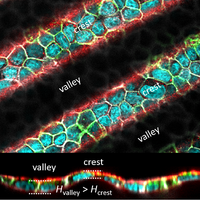
Epithelia are ubiquitous tissues that display a large diversity of functions and forms, from totally flat to highly curved. Various morphogenetic events, such as gastrulation or branching morphogenesis, correlate to changes in the curvature of epithelia. Building a physical framework to account for the shape of cells in epithelia is thus an important challenge to understand various normal and pathological biological processes, such as epithelial morphogenesis or cancer metastasis. It is widely recognized that the shape of epithelial cells is determined by the tension generated by the actomyosin cortex and the adhesion of cells to the substrate and to each other. These tensions and adhesions are not homogeneously distributed on the cell surface, which makes a 3D view of the problem valuable. To account for these biological and structural contributions to cell shape, different physical models have been proposed, which include surface energies, adhesions, line tensions, volume compressibility, or elasticity terms. However, an experimental procedure that would allow a validation of a minimal physical model for the shape of epithelial cells in 3D has not yet been proposed. In this study, we first made a quantitative analysis of the correlation between cell thickness and curvature during the formation of the ventral furrow in the early Drosophila embryo. We then cultured Madin-Darby Canine Kidney (MDCK) epithelial cells on substrates with a sinusoidal profile, allowing us to measure the shape of the cells on various positive and negative curvatures. We found that both in the early Drosophila ventral furrow and in MDCK epithelia cells are thicker when positively curved (on valleys of sinusoidal substrates) than when negatively curved (on the crests). The influence of curvature on the shape of epithelial cells could not be understood with a model using only differential apical, basal, and lateral surface energies. However, the addition of an apical line tension was sufficient to quantitatively account for the experimental measurements. The model also accounts for the shape of MDCK cells that overexpress E-cadherin. On the other hand, when reducing myosin II activity with blebbistatin, we measured a saturation of the difference in cell thickness between valleys and crests, suggesting the need for a term limiting large cell deformations. Our results show that a minimal model that accounts for epithelial cell shape needs to include an apical line tension in addition to differential surface energies, highlighting the importance of structures that produce anisotropic tension in epithelial cells, such as the actin belt linking adherens junctions. In the future, the model could be used to account for the shape of epithelial cells in different contexts, such as branching morphogenesis. Furthermore, our experimental procedure could be used to test a wider range of physical models for the shape of epithelia in curved environments, including, for example, continuous models.
中文翻译:

弯曲基质上上皮细胞的 3D 形状
上皮是无处不在的组织,其功能和形式多种多样,从完全平坦到高度弯曲。各种形态发生事件,如原肠胚形成或分支形态发生,与上皮曲率的变化相关。因此,建立一个物理框架来解释上皮细胞的形状是理解各种正常和病理生物学过程(例如上皮形态发生或癌症转移)的重要挑战。人们普遍认为,上皮细胞的形状是由肌动球蛋白皮层产生的张力以及细胞对基质和彼此的粘附决定的。这些张力和粘连并没有均匀地分布在细胞表面,这使得问题的 3D 视图很有价值。为了解释对细胞形状的这些生物学和结构贡献,已经提出了不同的物理模型,其中包括表面能、粘附、线张力、体积压缩率或弹性项。然而,尚未提出可以验证 3D 中上皮细胞形状的最小物理模型的实验程序。本研究首先定量分析了早期腹沟形成过程中细胞厚度与曲率的相关性。尚未提出可以验证 3D 中上皮细胞形状的最小物理模型的实验程序。本研究首先定量分析了早期腹沟形成过程中细胞厚度与曲率的相关性。尚未提出可以验证 3D 中上皮细胞形状的最小物理模型的实验程序。本研究首先定量分析了早期腹沟形成过程中细胞厚度与曲率的相关性。果蝇胚胎。然后,我们在具有正弦曲线的基底上培养了 Madin-Darby Canine Kidney (MDCK) 上皮细胞,使我们能够测量各种正曲率和负曲率的细胞形状。我们发现在早期的果蝇中腹侧沟和 MDCK 上皮细胞在正弯曲时(在正弦基底的谷部)比在负弯曲时(在波峰上)更厚。曲率对上皮细胞形状的影响无法通过仅使用不同的顶端、基底和侧表面能量的模型来理解。然而,增加顶端线张力足以定量解释实验测量。该模型还解释了过度表达 E-cadherin 的 MDCK 细胞的形状。另一方面,当用 blebbistatin 降低肌球蛋白 II 活性时,我们测量了谷和峰之间细胞厚度差异的饱和度,这表明需要限制大细胞变形的术语。我们的结果表明,解释上皮细胞形状的最小模型除了不同的表面能之外还需要包括顶端线张力,突出了在上皮细胞中产生各向异性张力的结构的重要性,例如连接粘附连接的肌动蛋白带。将来,该模型可用于解释不同背景下上皮细胞的形状,例如分支形态发生。此外,我们的实验程序可用于测试弯曲环境中上皮细胞形状的更广泛物理模型,包括例如连续模型。将来,该模型可用于解释不同背景下上皮细胞的形状,例如分支形态发生。此外,我们的实验程序可用于测试弯曲环境中上皮细胞形状的更广泛物理模型,包括例如连续模型。将来,该模型可用于解释不同背景下上皮细胞的形状,例如分支形态发生。此外,我们的实验程序可用于测试弯曲环境中上皮细胞形状的更广泛物理模型,包括例如连续模型。
更新日期:2021-08-04
中文翻译:
弯曲基质上上皮细胞的 3D 形状
上皮是无处不在的组织,其功能和形式多种多样,从完全平坦到高度弯曲。各种形态发生事件,如原肠胚形成或分支形态发生,与上皮曲率的变化相关。因此,建立一个物理框架来解释上皮细胞的形状是理解各种正常和病理生物学过程(例如上皮形态发生或癌症转移)的重要挑战。人们普遍认为,上皮细胞的形状是由肌动球蛋白皮层产生的张力以及细胞对基质和彼此的粘附决定的。这些张力和粘连并没有均匀地分布在细胞表面,这使得问题的 3D 视图很有价值。为了解释对细胞形状的这些生物学和结构贡献,已经提出了不同的物理模型,其中包括表面能、粘附、线张力、体积压缩率或弹性项。然而,尚未提出可以验证 3D 中上皮细胞形状的最小物理模型的实验程序。本研究首先定量分析了早期腹沟形成过程中细胞厚度与曲率的相关性。尚未提出可以验证 3D 中上皮细胞形状的最小物理模型的实验程序。本研究首先定量分析了早期腹沟形成过程中细胞厚度与曲率的相关性。尚未提出可以验证 3D 中上皮细胞形状的最小物理模型的实验程序。本研究首先定量分析了早期腹沟形成过程中细胞厚度与曲率的相关性。果蝇胚胎。然后,我们在具有正弦曲线的基底上培养了 Madin-Darby Canine Kidney (MDCK) 上皮细胞,使我们能够测量各种正曲率和负曲率的细胞形状。我们发现在早期的果蝇中腹侧沟和 MDCK 上皮细胞在正弯曲时(在正弦基底的谷部)比在负弯曲时(在波峰上)更厚。曲率对上皮细胞形状的影响无法通过仅使用不同的顶端、基底和侧表面能量的模型来理解。然而,增加顶端线张力足以定量解释实验测量。该模型还解释了过度表达 E-cadherin 的 MDCK 细胞的形状。另一方面,当用 blebbistatin 降低肌球蛋白 II 活性时,我们测量了谷和峰之间细胞厚度差异的饱和度,这表明需要限制大细胞变形的术语。我们的结果表明,解释上皮细胞形状的最小模型除了不同的表面能之外还需要包括顶端线张力,突出了在上皮细胞中产生各向异性张力的结构的重要性,例如连接粘附连接的肌动蛋白带。将来,该模型可用于解释不同背景下上皮细胞的形状,例如分支形态发生。此外,我们的实验程序可用于测试弯曲环境中上皮细胞形状的更广泛物理模型,包括例如连续模型。将来,该模型可用于解释不同背景下上皮细胞的形状,例如分支形态发生。此外,我们的实验程序可用于测试弯曲环境中上皮细胞形状的更广泛物理模型,包括例如连续模型。将来,该模型可用于解释不同背景下上皮细胞的形状,例如分支形态发生。此外,我们的实验程序可用于测试弯曲环境中上皮细胞形状的更广泛物理模型,包括例如连续模型。


 京公网安备 11010802027423号