Computers in Biology and Medicine ( IF 7.7 ) Pub Date : 2021-08-02 , DOI: 10.1016/j.compbiomed.2021.104721 Pankaj K Jain 1 , Neeraj Sharma 1 , Argiris A Giannopoulos 2 , Luca Saba 3 , Andrew Nicolaides 4 , Jasjit S Suri 5
|
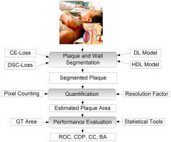
The automated and accurate carotid plaque segmentation in B-mode ultrasound (US) is an essential part of stroke risk stratification. Previous segmented methods used AtheroEdge™ 2.0 (AtheroPoint™, Roseville, CA) for the common carotid artery (CCA). This study focuses on automated plaque segmentation in the internal carotid artery (ICA) using solo deep learning (SDL) and hybrid deep learning (HDL) models.
The methodology consists of a novel design of 10 types of SDL/HDL models (AtheroEdge™ 3.0 systems (AtheroPoint™, Roseville, CA) with a depth of four layers each. Five of the models use cross-entropy (CE)-loss, and the other five models use Dice similarity coefficient (DSC)-loss functions derived from UNet, UNet+, SegNet, SegNet-UNet, and SegNet-UNet+. The K10 protocol (Train:Test:90%:10%) was applied for all 10 models for training and predicting (segmenting) the plaque region, which was then quantified to compute the plaque area in mm2. Further, the data augmentation effect was analyzed.
The database consisted of 970 ICA B-mode US scans taken from 99 moderate to high-risk patients. Using the difference area threshold of 10 mm2 between ground truth (GT) and artificial intelligence (AI), the area under the curve (AUC) values were 0.91, 0.911, 0.908, 0.905, and 0.898, all with a p-value of <0.001 (for CE-loss models) and 0.883, 0.889, 0.905, 0.889, and 0.907, all with a p-value of <0.001 (for DSC-loss models). The correlations between the AI-based plaque area and GT plaque area were 0.98, 0.96, 0.97, 0.98, and 0.97, all with a p-value of <0.001 (for CE-loss models) and 0.98, 0.98, 0.97, 0.98, and 0.98 (for DSC-loss models). Overall, the online system performs plaque segmentation in less than 1 s. We validate our hypothesis that HDL and SDL models demonstrate comparable performance. SegNet-UNet was the best-performing hybrid architecture.
中文翻译:
颈内动脉 B 型超声中动脉粥样硬化斑块的混合深度学习分割模型
B 型超声 (US) 中自动且准确的颈动脉斑块分割是中风风险分层的重要组成部分。以前的分段方法使用 AtheroEdge™ 2.0(AtheroPoint™,Roseville,CA)用于颈总动脉 (CCA)。本研究侧重于使用单独深度学习 (SDL) 和混合深度学习 (HDL) 模型在颈内动脉 (ICA) 中自动进行斑块分割。
该方法包括 10 种类型的 SDL/HDL 模型(AtheroEdge™ 3.0 系统(AtheroPoint™,Roseville,CA)的新颖设计,每层深度为四层。其中五个模型使用交叉熵 (CE)-损失,其他五个模型使用Dice相似系数(DSC)-损失函数,分别来自UNet、UNet+、SegNet、SegNet-UNet和SegNet-UNet+。K10协议(Train:Test:90%:10%)应用于所有10 个模型用于训练和预测(分割)斑块区域,然后对其进行量化以计算以 mm 2为单位的斑块面积。此外,分析了数据增强效果。
该数据库由来自99 名中高危患者的970 份ICA B 模式美国扫描组成。使用10毫米的差面积阈值2的地面实况(GT)和人工智能(AI),所述曲线下的面积之间(AUC)值分别为0.91,0.911,0.908,0.905,和0.898,都带有p的-值<0.001(对于 CE 损失模型)和0.883、0.889、0.905、0.889和0.907,均具有p-<0.001 的值(对于 DSC 损耗模型)。的基于AI的斑块面积和GT斑块区域之间的相关性是0.98,0.96,0.97,0.98,和0.97,所有的<0.001的p值(CE-损耗模型)和0.98,0.98,0.97,0.98,和0.98(对于 DSC 损耗模型)。总体而言,在线系统在不到 1 秒的时间内执行斑块分割。我们验证了我们的假设,即 HDL 和 SDL 模型表现出可比的性能。SegNet-UNet 是性能最好的混合架构。


 京公网安备 11010802027423号