Nanomedicine: Nanotechnology, Biology and Medicine ( IF 5.4 ) Pub Date : 2021-07-24 , DOI: 10.1016/j.nano.2021.102452 Eros Azzalini 1 , Nodira Abdurakhmanova 2 , Pietro Parisse 3 , Michele Bartoletti 4 , Vincenzo Canzonieri 1 , Giorgio Stanta 5 , Loredana Casalis 2 , Serena Bonin 5
|
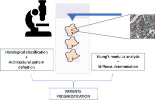
High grade serous ovarian carcinoma (HGSOC) is recognized as the most frequent type of ovarian cancer and the main cause of ovarian cancer related deaths worldwide. Although homologous recombination deficiency testing has been adopted in the clinical workflow, morphological analysis remains the main diagnostic tool. In this study Atomic Force Microscopy (AFM) was tested in standard hematoxylin and eosin (H&E) stained sections to investigate the biomechanical properties of different architectural growing patterns of HGSOC. Our results showed that AFM was able to discriminate HGSOC morphological growing patterns as well as patients’ stage. Micropapillary pattern, which has been associated to poor outcome, had lower Young’s moduli. In addition stage IV HGSOC was significantly softer than stage III cancers. Based on our results, AFM analysis could represent an additional tool in HGSOC morphological diagnosis as the biomechanical proprieties of HGSOC were quantitatively associated to tumor staging and architectural pattern.
中文翻译:
高级别浆液性卵巢癌临床样本中的细胞硬度和形态结构模式
高级别浆液性卵巢癌(HGSOC)被认为是最常见的卵巢癌类型,也是全世界卵巢癌相关死亡的主要原因。尽管在临床工作流程中已采用同源重组缺陷检测,但形态分析仍然是主要的诊断工具。在这项研究中,原子力显微镜 (AFM) 在标准苏木精和伊红 (H&E) 染色切片中进行了测试,以研究 HGSOC 不同建筑生长模式的生物力学特性。我们的结果表明,AFM 能够区分 HGSOC 形态生长模式以及患者的阶段。与不良结果相关的微毛细管模式具有较低的杨氏模量。此外,IV 期 HGSOC 明显比 III 期癌症软。根据我们的结果,


 京公网安备 11010802027423号