当前位置:
X-MOL 学术
›
Brain Pathol.
›
论文详情
Our official English website, www.x-mol.net, welcomes your feedback! (Note: you will need to create a separate account there.)
Microglia activation in postmortem brains with schizophrenia demonstrates distinct morphological changes between brain regions
Brain Pathology ( IF 6.4 ) Pub Date : 2021-07-23 , DOI: 10.1111/bpa.13003 Ryan Gober 1 , Maryam Ardalan 2, 3 , Seyedeh Marziyeh Jabbari Shiadeh 2, 3 , Linda Duque 1 , Susanna P Garamszegi 1 , Maureen Ascona 1 , Ayled Barreda 1 , Xiaoyan Sun 1 , Carina Mallard 2 , Regina T Vontell 1
Brain Pathology ( IF 6.4 ) Pub Date : 2021-07-23 , DOI: 10.1111/bpa.13003 Ryan Gober 1 , Maryam Ardalan 2, 3 , Seyedeh Marziyeh Jabbari Shiadeh 2, 3 , Linda Duque 1 , Susanna P Garamszegi 1 , Maureen Ascona 1 , Ayled Barreda 1 , Xiaoyan Sun 1 , Carina Mallard 2 , Regina T Vontell 1
Affiliation
|
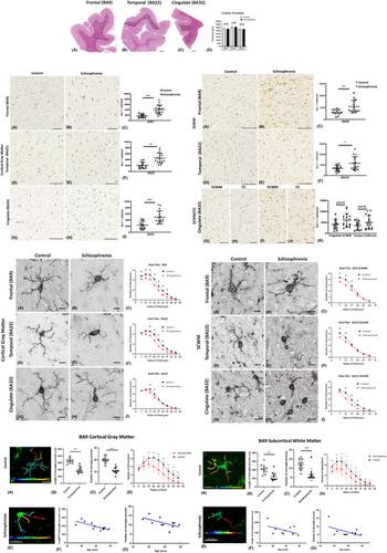
Schizophrenia (SCZ) is a psychiatric disorder that can include symptoms of disorganized speech and thoughts with uncertain underlying mechanisms possibly linked to over-activated microglia. In this study, we used brain samples from sixteen donors with SCZ and thirteen control donors to assess the differential activation of microglia by quantifying density and 3D reconstruction of microglia stained with ionized calcium-binding adaptor molecule-1 (Iba1). Our samples consisted of sections from the frontal, temporal, and cingulate cortical gray matter, subcortical white matter regions (SCWM), and included the anterior corpus callosum. In the first series of studies, we performed a density analysis followed by a spatial analysis to ascertain the microglial density, distribution, and soma size in SCZ brains. Second, we performed a series of morphological quantification techniques to investigate the arborization patterns of the microglia in SCZ. The results demonstrated an increase in microglia density in the cortical gray matter regions in SCZ cases, while in the SCWM, there was a significant increase in microglia density in the frontal and temporal, but not in the other brain regions of interest (ROIs). Spatial analysis using the “nearest neighbor” demonstrated that there was no effect in “clustering”, but there were shorter distances between microglia seen in the SCZ cases. The morphological measures showed that there was a region-dependent increase in the microglia soma size in the SCZ cases while the Sholl analysis revealed a significant decrease in the microglia arborization in the SCZ cases across all the ROI’s studied. An in-depth 3D reconstruction of microglia in Brodmann area 9 cortical region found that there was a significant association between age and reduced microglial arborization in the SCZ cases. This region-dependent age association can help determine whether longitudinal changes in microglial activation across age are brain region-dependent, which may point to potential therapeutic targets.
中文翻译:

精神分裂症死后大脑中的小胶质细胞激活显示大脑区域之间存在明显的形态变化
精神分裂症 (SCZ) 是一种精神疾病,可能包括言语和思想杂乱无章的症状,其潜在机制不确定,可能与过度激活的小胶质细胞有关。在这项研究中,我们使用来自 16 名 SCZ 供体和 13 名对照供体的脑样本,通过量化用离子化钙结合接头分子-1 (Iba1) 染色的小胶质细胞的密度和 3D 重建来评估小胶质细胞的差异激活。我们的样本包括来自额叶、颞叶和扣带皮层灰质、皮层下白质区域 (SCWM) 的部分,并包括前胼胝体。在第一系列研究中,我们进行了密度分析,然后进行了空间分析,以确定 SCZ 大脑中的小胶质细胞密度、分布和体细胞大小。第二,我们进行了一系列形态学量化技术来研究 SCZ 中小胶质细胞的树枝状模式。结果表明,在 SCZ 病例中,皮质灰质区域的小胶质细胞密度增加,而在 SCWM 中,额叶和颞叶的小胶质细胞密度显着增加,但在其他大脑感兴趣区域 (ROI) 中没有。使用“最近邻”的空间分析表明,“聚类”没有影响,但在 SCZ 病例中看到的小胶质细胞之间的距离较短。形态学测量表明,SCZ 病例中的小胶质细胞体大小呈区域依赖性增加,而 Sholl 分析显示,在所有研究的 ROI 中,SCZ 病例中的小胶质细胞树突显着减少。对 Brodmann 9 区皮层区域小胶质细胞的深入 3D 重建发现,SCZ 病例中年龄与小胶质细胞树枝状结构减少之间存在显着关联。这种依赖于区域的年龄关联可以帮助确定跨年龄小胶质细胞激活的纵向变化是否依赖于大脑区域,这可能指向潜在的治疗目标。
更新日期:2021-07-23
中文翻译:
精神分裂症死后大脑中的小胶质细胞激活显示大脑区域之间存在明显的形态变化
精神分裂症 (SCZ) 是一种精神疾病,可能包括言语和思想杂乱无章的症状,其潜在机制不确定,可能与过度激活的小胶质细胞有关。在这项研究中,我们使用来自 16 名 SCZ 供体和 13 名对照供体的脑样本,通过量化用离子化钙结合接头分子-1 (Iba1) 染色的小胶质细胞的密度和 3D 重建来评估小胶质细胞的差异激活。我们的样本包括来自额叶、颞叶和扣带皮层灰质、皮层下白质区域 (SCWM) 的部分,并包括前胼胝体。在第一系列研究中,我们进行了密度分析,然后进行了空间分析,以确定 SCZ 大脑中的小胶质细胞密度、分布和体细胞大小。第二,我们进行了一系列形态学量化技术来研究 SCZ 中小胶质细胞的树枝状模式。结果表明,在 SCZ 病例中,皮质灰质区域的小胶质细胞密度增加,而在 SCWM 中,额叶和颞叶的小胶质细胞密度显着增加,但在其他大脑感兴趣区域 (ROI) 中没有。使用“最近邻”的空间分析表明,“聚类”没有影响,但在 SCZ 病例中看到的小胶质细胞之间的距离较短。形态学测量表明,SCZ 病例中的小胶质细胞体大小呈区域依赖性增加,而 Sholl 分析显示,在所有研究的 ROI 中,SCZ 病例中的小胶质细胞树突显着减少。对 Brodmann 9 区皮层区域小胶质细胞的深入 3D 重建发现,SCZ 病例中年龄与小胶质细胞树枝状结构减少之间存在显着关联。这种依赖于区域的年龄关联可以帮助确定跨年龄小胶质细胞激活的纵向变化是否依赖于大脑区域,这可能指向潜在的治疗目标。


 京公网安备 11010802027423号