NeuroImage: Clinical ( IF 4.2 ) Pub Date : 2021-07-14 , DOI: 10.1016/j.nicl.2021.102756 Jérôme Baranger 1 , Olivier Villemain 2 , Matthias Wagner 3 , Mariella Vargas-Gutierrez 4 , Mike Seed 5 , Olivier Baud 6 , Birgit Ertl-Wagner 3 , Julien Aguet 7
|
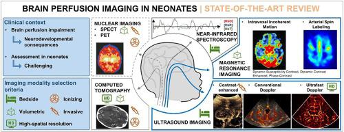
Abnormal variations of the neonatal brain perfusion can result in long-term neurodevelopmental consequences and cerebral perfusion imaging can play an important role in diagnostic and therapeutic decision-making. To identify at-risk situations, perfusion imaging of the neonatal brain must accurately evaluate both regional and global perfusion. To date, neonatal cerebral perfusion assessment remains challenging. The available modalities such as magnetic resonance imaging (MRI), ultrasound imaging, computed tomography (CT), near-infrared spectroscopy or nuclear imaging have multiple compromises and limitations. Several promising methods are being developed to achieve better diagnostic accuracy and higher robustness, in particular using advanced MRI and ultrasound techniques.
The objective of this state-of-the-art review is to analyze the methodology and challenges of neonatal brain perfusion imaging, to describe the currently available modalities, and to outline future perspectives.
中文翻译:
新生儿脑灌注成像
新生儿脑灌注的异常变化会导致长期的神经发育后果,而脑灌注成像可以在诊断和治疗决策中发挥重要作用。为了识别有风险的情况,新生儿大脑的灌注成像必须准确评估区域和全局灌注。迄今为止,新生儿脑灌注评估仍然具有挑战性。可用的模式,例如磁共振成像 (MRI)、超声成像、计算机断层扫描 (CT)、近红外光谱或核成像,具有多种折衷和局限性。正在开发几种有前途的方法来实现更好的诊断准确性和更高的鲁棒性,特别是使用先进的 MRI 和超声技术。
这篇最新综述的目的是分析新生儿脑灌注成像的方法和挑战,描述当前可用的模式,并概述未来的前景。


 京公网安备 11010802027423号