当前位置:
X-MOL 学术
›
Luminescence
›
论文详情
Our official English website, www.x-mol.net, welcomes your feedback! (Note: you will need to create a separate account there.)
Spectroscopic analysis of coenzyme binding to betaine aldehyde dehydrogenase dependent on potassium
Luminescence ( IF 2.9 ) Pub Date : 2021-07-02 , DOI: 10.1002/bio.4115 César Muñoz-Bacasehua 1 , Jesús A Rosas-Rodríguez 2 , Alexis Alonso López-Zavala 3 , Elisa M Valenzuela-Soto 1
Luminescence ( IF 2.9 ) Pub Date : 2021-07-02 , DOI: 10.1002/bio.4115 César Muñoz-Bacasehua 1 , Jesús A Rosas-Rodríguez 2 , Alexis Alonso López-Zavala 3 , Elisa M Valenzuela-Soto 1
Affiliation
|
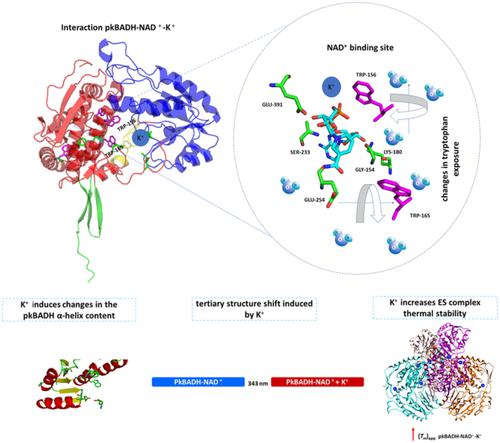
Glycine betaine is the main osmolyte synthesized and accumulated in mammalian renal cells. Glycine betaine synthesis is catalyzed by the enzyme betaine aldehyde dehydrogenase (BADH) using NAD+ as the coenzyme. Previous studies have shown that porcine kidney betaine aldehyde dehydrogenase (pkBADH) binds NAD+ with different affinities at each active site and that the binding is K+ dependent. The objective of this work was to analyze the changes in the pkBADH secondary and tertiary structure resulting from variable concentrations of NAD+ and the role played by K+. Intrinsic fluorescence studies were carried out at fixed-variable concentrations of K+ and titrating the enzyme with varying concentrations of NAD+. Fluorescence analysis showed a shift of the maximum emission towards red as the concentration of K+ was increased. Changes in the exposure of tryptophan located near the NAD+ binding site were found when the enzyme was titrated with NAD+ in the presence of potassium. Fluorescence data analysis showed that the K+ presence promoted static quenching that facilitated the pkBADH–NAD+ complex formation. DC data analysis showed that binding of K+ to the enzyme caused changes in the α-helix content of 4% and 12% in the presence of 25 mM and 100 mM K+, respectively. The presence of K+ during NAD+ binding to pkBADH increased the thermal stability of the complex. These results indicated that K+ facilitated the pkBADH–NAD+ complex formation and suggested that K+ caused small changes in secondary and tertiary structures that could influence the active site conformation.
中文翻译:

辅酶结合依赖钾的甜菜碱醛脱氢酶的光谱分析
甘氨酸甜菜碱是哺乳动物肾细胞中合成和积累的主要渗透剂。甘氨酸甜菜碱合成由甜菜碱醛脱氢酶 (BADH) 催化,使用 NAD +作为辅酶。先前的研究表明,猪肾甜菜碱醛脱氢酶 (pkBADH)在每个活性位点以不同的亲和力结合 NAD +并且这种结合是 K +依赖性的。这项工作的目的是分析由不同浓度的 NAD +引起的 pkBADH 二级和三级结构的变化以及 K + 所起的作用。在固定可变浓度的 K + 下进行本征荧光研究并用不同浓度的 NAD +滴定酶。荧光分析表明,随着 K +浓度的增加,最大发射向红色移动。在存在钾的情况下用 NAD +滴定酶时,发现位于 NAD +结合位点附近的色氨酸暴露发生变化。荧光数据分析表明,K + 的存在促进了静态猝灭,从而促进了 pkBADH-NAD +复合物的形成。DC数据分析表明,在25 mM和100 mM K +存在下,K +与酶的结合导致α-螺旋含量发生4%和12%的变化, 分别。NAD +与 pkBADH 结合期间K +的存在增加了复合物的热稳定性。这些结果表明 K +促进了 pkBADH-NAD +复合物的形成,并表明 K +引起二级和三级结构的微小变化,这可能会影响活性位点构象。
更新日期:2021-07-02
中文翻译:
辅酶结合依赖钾的甜菜碱醛脱氢酶的光谱分析
甘氨酸甜菜碱是哺乳动物肾细胞中合成和积累的主要渗透剂。甘氨酸甜菜碱合成由甜菜碱醛脱氢酶 (BADH) 催化,使用 NAD +作为辅酶。先前的研究表明,猪肾甜菜碱醛脱氢酶 (pkBADH)在每个活性位点以不同的亲和力结合 NAD +并且这种结合是 K +依赖性的。这项工作的目的是分析由不同浓度的 NAD +引起的 pkBADH 二级和三级结构的变化以及 K + 所起的作用。在固定可变浓度的 K + 下进行本征荧光研究并用不同浓度的 NAD +滴定酶。荧光分析表明,随着 K +浓度的增加,最大发射向红色移动。在存在钾的情况下用 NAD +滴定酶时,发现位于 NAD +结合位点附近的色氨酸暴露发生变化。荧光数据分析表明,K + 的存在促进了静态猝灭,从而促进了 pkBADH-NAD +复合物的形成。DC数据分析表明,在25 mM和100 mM K +存在下,K +与酶的结合导致α-螺旋含量发生4%和12%的变化, 分别。NAD +与 pkBADH 结合期间K +的存在增加了复合物的热稳定性。这些结果表明 K +促进了 pkBADH-NAD +复合物的形成,并表明 K +引起二级和三级结构的微小变化,这可能会影响活性位点构象。


 京公网安备 11010802027423号