当前位置:
X-MOL 学术
›
Brain Pathol.
›
论文详情
Our official English website, www.x-mol.net, welcomes your feedback! (Note: you will need to create a separate account there.)
Role of VAPB and vesicular profiles in α-synuclein aggregates in multiple system atrophy
Brain Pathology ( IF 6.4 ) Pub Date : 2021-07-01 , DOI: 10.1111/bpa.13001 Fumiaki Mori 1 , Yasuo Miki 1 , Kunikazu Tanji 1 , Tomoya Kon 2 , Masahiko Tomiyama 2 , Akiyoshi Kakita 3 , Koichi Wakabayashi 1
Brain Pathology ( IF 6.4 ) Pub Date : 2021-07-01 , DOI: 10.1111/bpa.13001 Fumiaki Mori 1 , Yasuo Miki 1 , Kunikazu Tanji 1 , Tomoya Kon 2 , Masahiko Tomiyama 2 , Akiyoshi Kakita 3 , Koichi Wakabayashi 1
Affiliation
|
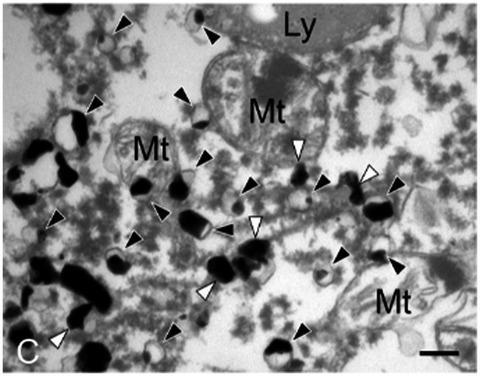
The pathological hallmark of multiple system atrophy (MSA) is fibrillary aggregates of α-synuclein (α-Syn) in the cytoplasm and nucleus of both oligodendrocytes and neurons. In neurons, α-Syn localizes to the cytosolic and membrane compartments, including the synaptic vesicles, mitochondria, and endoplasmic reticulum (ER). α-Syn binds to vesicle-associated membrane protein-binding protein B (VAPB) in the ER membrane. Overexpression of wild-type and familial Parkinson's disease mutant α-Syn perturbs the association between the ER and mitochondria, leading to ER stress and ultimately neurodegeneration. We examined brains from MSA patients (n = 7) and control subjects (n = 5) using immunohistochemistry and immunoelectron microscopy with antibodies against VAPB and phosphorylated α-Syn. In controls, the cytoplasm of neurons and glial cells was positive for VAPB, whereas in MSA lesions VAPB immunoreactivity was decreased. The proportion of VAPB-negative neurons in the pontine nucleus was significantly higher in MSA (13.6%) than in controls (0.6%). The incidence of cytoplasmic inclusions in VAPB-negative neurons was significantly higher (42.2%) than that in VAPB-positive neurons (3.6%); 67.2% of inclusion-bearing oligodendrocytes and 51.1% of inclusion-containing neurons were negative for VAPB. Immunoelectron microscopy revealed that α-Syn and VAPB were localized to granulofilamentous structures in the cytoplasm of oligodendrocytes and neurons. Many vesicular structures labeled with anti-α-Syn were also observed within the granulofilamentous structures in the cytoplasm and nucleus of both oligodendrocytes and neurons. These findings suggest that, in MSA, reduction of VAPB is involved in the disease process and that vesicular structures are associated with inclusion formation.
中文翻译:

VAPB 和囊泡分布在多系统萎缩中 α-突触核蛋白聚集体中的作用
多系统萎缩 (MSA) 的病理标志是少突胶质细胞和神经元的细胞质和细胞核中的 α-突触核蛋白 (α-Syn) 纤维状聚集体。在神经元中,α-Syn 定位于胞质和膜区室,包括突触小泡、线粒体和内质网 (ER)。α-Syn 与 ER 膜中的囊泡相关膜蛋白结合蛋白 B (VAPB) 结合。野生型和家族性帕金森病突变体 α-Syn 的过表达扰乱了 ER 和线粒体之间的关联,导致 ER 应激并最终导致神经变性。我们使用免疫组织化学和免疫电子显微镜检查了 MSA 患者 (n = 7) 和对照受试者 (n = 5) 的大脑,其中含有针对 VAPB 和磷酸化 α-Syn 的抗体。在控件中,神经元和神经胶质细胞的细胞质对 VAPB 呈阳性,而在 MSA 病变中 VAPB 免疫反应性降低。MSA组(13.6%)脑桥核中VAPB阴性神经元的比例显着高于对照组(0.6%)。VAPB 阴性神经元胞质包涵体的发生率(42.2%)显着高于 VAPB 阳性神经元(3.6%);67.2% 的含有包涵体的少突胶质细胞和 51.1% 的含有包涵体的神经元对 VAPB 呈阴性。免疫电镜显示 α-Syn 和 VAPB 定位于少突胶质细胞和神经元细胞质中的粒丝状结构。在少突胶质细胞和神经元的细胞质和细胞核中的粒丝状结构中也观察到许多用抗α-Syn标记的囊泡结构。
更新日期:2021-07-01
中文翻译:
VAPB 和囊泡分布在多系统萎缩中 α-突触核蛋白聚集体中的作用
多系统萎缩 (MSA) 的病理标志是少突胶质细胞和神经元的细胞质和细胞核中的 α-突触核蛋白 (α-Syn) 纤维状聚集体。在神经元中,α-Syn 定位于胞质和膜区室,包括突触小泡、线粒体和内质网 (ER)。α-Syn 与 ER 膜中的囊泡相关膜蛋白结合蛋白 B (VAPB) 结合。野生型和家族性帕金森病突变体 α-Syn 的过表达扰乱了 ER 和线粒体之间的关联,导致 ER 应激并最终导致神经变性。我们使用免疫组织化学和免疫电子显微镜检查了 MSA 患者 (n = 7) 和对照受试者 (n = 5) 的大脑,其中含有针对 VAPB 和磷酸化 α-Syn 的抗体。在控件中,神经元和神经胶质细胞的细胞质对 VAPB 呈阳性,而在 MSA 病变中 VAPB 免疫反应性降低。MSA组(13.6%)脑桥核中VAPB阴性神经元的比例显着高于对照组(0.6%)。VAPB 阴性神经元胞质包涵体的发生率(42.2%)显着高于 VAPB 阳性神经元(3.6%);67.2% 的含有包涵体的少突胶质细胞和 51.1% 的含有包涵体的神经元对 VAPB 呈阴性。免疫电镜显示 α-Syn 和 VAPB 定位于少突胶质细胞和神经元细胞质中的粒丝状结构。在少突胶质细胞和神经元的细胞质和细胞核中的粒丝状结构中也观察到许多用抗α-Syn标记的囊泡结构。


 京公网安备 11010802027423号