Vibrational Spectroscopy ( IF 2.5 ) Pub Date : 2021-05-21 , DOI: 10.1016/j.vibspec.2021.103260 Tian Ning , Heping Li , Yishen Chen , Baoping Zhang , Furong Zhang , Shuang Wang
|
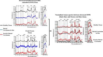
The application of Raman microspectroscopy in the histopathological analysis have been widely pursued for characterizing ex vivo biological tissue, and for reducing the number of false positive biopsies and increase the cancer diagnosis accuracy. Many studies have been carried out on fresh or frozen tissue samples, which preserved comprehensive qualitative and quantitative compositional information. However, by the wider applicability of dewaxed formalin-fixed paraffin-embedded (FFPE) samples in the hospital, an abundant tissue bank is a useful resource for the on-site retrospective research to improve diagnosis accuracy. Therefore, we implemented an ex vivo Raman spectroscopic study combined multivariate analysis methods for presenting a better understanding of the sample processing effects and testifying the potential Raman based pathological analysis capabilities of FFPE sections. By comparing 600 spectra from thirty-six fresh frozen (n=18) and FFPE breast tissue samples (n=18), including Healthy tissue, Ductal Carcinoma In Situ, and Invasive Ductal Carcinoma tissue, the results demonstrated that the dewaxing process significantly altered the biochemical composition of the tissues, particularly lipids, proteins, and carotenoids. Even though, the analytical result proved that commonly used multivariate analysis methods, including principal component analysis - linear discriminant analysis (PCA-LDA) and partial least squares- discriminant analysis (PLS-DA), could still distinguish the investigated tissue types effectively with satisfying overall accuracy in which PCA-LDA is 88.3 %, PLS-DA is 93.0 %. Therefore, this study confirmed that FFPE sections have diagnostic potential with multivariate analytical model provided that the biochemical changes, meanwhile tissue processing should be aware.
中文翻译:
基于拉曼光谱的福尔马林固定石蜡包埋的乳腺癌组织的病理分析和鉴别
拉曼光谱法在组织病理学分析中的应用已被广泛追求以表征离体生物组织,并减少假阳性活组织检查的数量并提高癌症诊断的准确性。已经对新鲜或冷冻的组织样本进行了许多研究,这些样本保留了全面的定性和定量组成信息。然而,由于脱蜡的福尔马林固定石蜡包埋(FFPE)样品在医院中的广泛应用,丰富的组织库是用于现场回顾性研究以提高诊断准确性的有用资源。因此,我们实施了离体拉曼光谱研究结合了多元分析方法,以更好地理解样品的处理效果,并证明了FFPE切片潜在的基于拉曼的病理分析能力。通过比较36种新鲜冷冻(n = 18)和FFPE乳腺组织样本(n = 18)的600光谱,包括健康组织,导管原位癌和浸润性导管癌组织,结果表明脱蜡过程显着改变了组织的生化成分,特别是脂质,蛋白质和类胡萝卜素。尽管分析结果证明了常用的多元分析方法,包括主成分分析-线性判别分析(PCA-LDA)和偏最小二乘-判别分析(PLS-DA),PCA-LDA为88.3%,PLS-DA为93.0%,仍能以令人满意的整体精度有效地区分所研究的组织类型。因此,本研究证实,只要生化改变,FFPE切片具有多变量分析模型的诊断潜力,同时应注意组织处理。


 京公网安备 11010802027423号