Journal of Structural Biology ( IF 3 ) Pub Date : 2021-05-16 , DOI: 10.1016/j.jsb.2021.107746 Jennifer Jiang 1 , Kuan Yu Cheong 2 , Paul G Falkowski 3 , Wei Dai 1
|
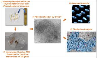
A long-standing challenge in cell biology is elucidating the structure and spatial distribution of individual membrane-bound proteins, protein complexes and their interactions in their native environment. Here, we describe a workflow that combines on-grid immunogold labeling, followed by cryo-electron tomography (cryoET) imaging and structural analyses to identify and characterize the structure of photosystem II (PSII) complexes. Using an antibody specific to a core subunit of PSII, the D1 protein (uniquely found in the water splitting complex in all oxygenic photoautotrophs), we identified PSII complexes in biophysically active thylakoid membranes isolated from a model marine diatom Phaeodactylum tricornutum. Subsequent cryoET analyses of these protein complexes resolved two PSII structures: supercomplexes and dimeric cores. Our integrative approach establishes the structural signature of multimeric membrane protein complexes in their native environment and provides a pathway to elucidate their high-resolution structures.
中文翻译:
集成网格免疫金标记和冷冻电子断层扫描揭示类囊体膜中的光系统 II 结构和空间分布
细胞生物学的一个长期挑战是阐明单个膜结合蛋白、蛋白复合物的结构和空间分布及其在天然环境中的相互作用。在这里,我们描述了一个工作流程,该工作流程结合了网格免疫金标记,然后进行冷冻电子断层扫描 (cryoET) 成像和结构分析,以识别和表征光系统 II (PSII) 复合物的结构。使用针对 PSII 核心亚基 D1 蛋白(唯一存在于所有含氧光自养生物的水分解复合物中)的抗体,我们在从模型海洋硅藻三角褐指藻中分离出的具有生物物理活性的类囊体膜中鉴定出了 PSII 复合物。随后对这些蛋白质复合物进行冷冻电子显微镜分析,解析了两种 PSII 结构:超复合物和二聚体核心。我们的综合方法建立了多聚膜蛋白复合物在其天然环境中的结构特征,并提供了阐明其高分辨率结构的途径。


 京公网安备 11010802027423号