Computers in Biology and Medicine ( IF 7.7 ) Pub Date : 2021-05-02 , DOI: 10.1016/j.compbiomed.2021.104454 Pradeep Kumar Chaudhary 1 , Ram Bilas Pachori 1
|
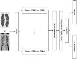
This work introduces the Fourier-Bessel series expansion-based decomposition (FBSED) method, which is an implementation of the wavelet packet decomposition approach in the Fourier-Bessel series expansion domain. The proposed method has been used for the diagnosis of pneumonia caused by the 2019 novel coronavirus disease (COVID-19) using chest X-ray image (CXI) and chest computer tomography image (CCTI). The FBSED method is used to decompose CXI and CCTI into sub-band images (SBIs). The SBIs are then used to train various pre-trained convolutional neural network (CNN) models separately using a transfer learning approach. The combination of SBI and CNN is termed as one channel. Deep features from each channel are fused to get a feature vector. Different classifiers are used to classify pneumonia caused by COVID-19 from other viral and bacterial pneumonia and healthy subjects with the extracted feature vector. The different combinations of channels have also been analyzed to make the process computationally efficient. For CXI and CCTI databases, the best performance has been obtained with only one and four channels, respectively. The proposed model was evaluated using 5-fold and 10-fold cross-validation processes. The average accuracy for the CXI database was 100% for both 5-fold and 10-fold cross-validation processes, and for the CCTI database, it is 97.6% for the 5-fold cross-validation process. Therefore, the proposed method may be used by radiologists to rapidly diagnose patients with COVID-19.
中文翻译:
使用X射线和CT图像基于FBSED的COVID-19自动诊断
本文介绍了基于傅立叶-贝塞尔级数展开的分解(FBSED)方法,该方法是在傅立叶-贝塞尔级数展开域中小波包分解方法的实现。该方法已通过胸部X射线图像(CXI)和胸部计算机断层扫描图像(CCTI)用于诊断由2019年新型冠状病毒病(COVID-19)引起的肺炎。FBSED方法用于将CXI和CCTI分解为子带图像(SBI)。然后,将SBI用于使用转移学习方法分别训练各种预训练的卷积神经网络(CNN)模型。SBI和CNN的组合称为一个通道。将来自每个通道的深层特征融合以获得特征向量。使用不同的分类器,使用提取的特征向量,将其他病毒性和细菌性肺炎以及健康人中由COVID-19引起的肺炎分类。还对通道的不同组合进行了分析,以使过程的计算效率更高。对于CXI和CCTI数据库,分别仅通过一个和四个通道即可获得最佳性能。使用5倍和10倍交叉验证过程对提出的模型进行了评估。CXI数据库的5倍和10倍交叉验证过程的平均准确度均为100%,而CCTI数据库的5倍交叉验证过程的平均准确度为97.6%。因此,放射科医生可以使用所提出的方法快速诊断COVID-19患者。还对通道的不同组合进行了分析,以使过程的计算效率更高。对于CXI和CCTI数据库,分别仅通过一个和四个通道即可获得最佳性能。使用5倍和10倍交叉验证过程对提出的模型进行了评估。CXI数据库的5倍和10倍交叉验证过程的平均准确度均为100%,而CCTI数据库的5倍交叉验证过程的平均准确度为97.6%。因此,放射科医生可以使用所提出的方法快速诊断COVID-19患者。还对通道的不同组合进行了分析,以使过程的计算效率更高。对于CXI和CCTI数据库,分别仅通过一个和四个通道即可获得最佳性能。使用5倍和10倍交叉验证过程对提出的模型进行了评估。CXI数据库的5倍和10倍交叉验证过程的平均准确度均为100%,而CCTI数据库的5倍交叉验证过程的平均准确度为97.6%。因此,放射科医生可以使用所提出的方法快速诊断COVID-19患者。使用5倍和10倍交叉验证过程对提出的模型进行了评估。CXI数据库的5倍和10倍交叉验证过程的平均准确度均为100%,而CCTI数据库的5倍交叉验证过程的平均准确度为97.6%。因此,放射科医生可以使用所提出的方法快速诊断COVID-19患者。使用5倍和10倍交叉验证过程对提出的模型进行了评估。CXI数据库的5倍和10倍交叉验证过程的平均准确度均为100%,而CCTI数据库的5倍交叉验证过程的平均准确度为97.6%。因此,放射科医生可以使用所提出的方法快速诊断COVID-19患者。


 京公网安备 11010802027423号