Signal Processing: Image Communication ( IF 3.5 ) Pub Date : 2021-05-03 , DOI: 10.1016/j.image.2021.116303 Huaifei Hu , Ning Pan , Haihua Liu , Liman Liu , Tailang Yin , Zhigang Tu , Alejandro F. Frangi
|
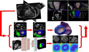
Segmentation of the left and right ventricles in cardiac MRI (Magnetic Resonance Imaging) is a prerequisite step for evaluating global and regional cardiac function. This work presents a novel and robust schema for MRI segmentation by combining the advantages of deep learning localization and 3D-ASM (3D Active Shape Model) restriction without any user interaction. Three fundamental techniques are exploited: (1) manual 2D contours are used to build distance maps to get 3D ground truth shape, (2) derived right ventricle points are employed to rotate the coarse initial shape for a refined bi-ventricle initial estimation, (3) segmentation results from deep learning are utilised to build distance maps for the 3D-ASM matching process to help image intensity modelling. The datasets used for experimenting the cine MRI data are 1000 cases from UK Biobank, 500 subjects are selected to train CNN (Convolution Neural Network) parameters, and the remaining 500 cases are adopted for validation. Specifically, cases are used to rebuild point distribution and image intensity models, and also utilized to train CNN. In addition, the left 500 cases are used to perform the validation experiments. For the segmentation of the RV (Right Ventricle) endocardial contour, LV (Left Ventricle) endo- and epicardial contours, overlap, Jaccard similarity index, Point-to-surface errors and cardiac functional parameters are calculated. Experimental results show that the proposed method has advantages over the previous approaches.
中文翻译:
使用3D-ASM和深度学习在心脏MRI中自动分割左右心室
在心脏MRI(磁共振成像)中对左心室和右心室进行分割是评估整体和局部心脏功能的先决条件。这项工作结合了深度学习定位和3D-ASM(3D活动形状模型)限制的优势,而无需任何用户交互,从而为MRI分割提供了一种新颖而强大的方案。利用了三种基本技术:(1)使用手动2D轮廓线建立距离图以获得3D地面真值形状;(2)使用派生的右心室点旋转粗略的初始形状,以进行精确的双心室初始估计,( 3)深度学习的分割结果用于为3D-ASM匹配过程建立距离图,以帮助进行图像强度建模。用于电影MRI数据实验的数据集来自UK Biobank,1000例,选择了500个主题来训练CNN(卷积神经网络)参数,并采用其余500个案例进行验证。具体来说,案例用于重建点分布和图像强度模型,还用于训练CNN。此外,剩下的500个案例用于执行验证实验。对于RV(右心室)心内膜轮廓,LV(左心室)心内膜和心外膜轮廓的分割,计算重叠度,Jaccard相似性指数,点对面误差和心脏功能参数。实验结果表明,该方法具有优于现有方法的优点。案例用于重建点分布和图像强度模型,还用于训练CNN。此外,剩下的500个案例用于执行验证实验。对于RV(右心室)心内膜轮廓,LV(左心室)心内膜和心外膜轮廓的分割,计算重叠度,Jaccard相似性指数,点对面误差和心脏功能参数。实验结果表明,该方法具有优于现有方法的优点。案例用于重建点分布和图像强度模型,还用于训练CNN。此外,剩下的500个案例用于执行验证实验。对于RV(右心室)心内膜轮廓,LV(左心室)心内膜和心外膜轮廓的分割,计算重叠度,Jaccard相似性指数,点对面误差和心脏功能参数。实验结果表明,该方法具有优于现有方法的优点。计算点到面误差和心脏功能参数。实验结果表明,该方法具有优于现有方法的优点。计算点到面误差和心脏功能参数。实验结果表明,该方法具有优于现有方法的优点。


 京公网安备 11010802027423号