Polymer Bulletin ( IF 3.2 ) Pub Date : 2021-04-19 , DOI: 10.1007/s00289-021-03702-0 Parviz Ranjbarvan , Ali Golchin , Arezo Azari , Zahra Niknam
|
Abstract
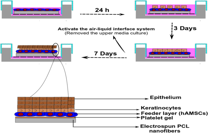
In the present study, neonate keratinocytes were cocultured with human adipose-derived mesenchymal stem cells (hAMSCs) on the electrospun polycaprolactone-platelet gel (PCL–PG) scaffold. To evaluate its potential of wound healing and skin tissue engineering, neonate keratinocytes must be differentiated. The PCL scaffold prepared by the electrospinning technique was fabricated and coated by PG. Then scaffolds were fully characterized by SEM, contact angle, FTIR–ATR, and tensile test. Following seeding hAMSCs as a feeder layer, neonate keratinocytes were cocultured directly on PCL–PG scaffolds and neat PCL. The hAMSCs and neonate keratinocytes' viability was measured using MTT assay for up to 10 days (1st, 3rd, 5th, 7th, and 10th days). To examine the epidermal maturation, cytokeratin 10 and loricrin determinants detection was used through immunocytochemistry (ICC). In addition, real-time PCR was used to examine keratinocyte marker genes in keratin 10, keratin 14, and Involucrin. The MTT assay results showed higher cell viability and the proliferation of neo-keratinocytes on PCL–PG fibrillar scaffolds than the PCL scaffold. On the PCL–PG scaffolds in the presence of hAMSCs as a feeder layer, the PCR and ICC analysis had a higher cell differentiation compared to neat PCL scaffolds. Moreover, SEM images showed that the keratinocytes cocultured with hAMSCs demonstrated a better proliferation and adhesion on PCL–PG nanofiber scaffolds. Based on results, PG increased the nanofibrous PCL scaffold's biological properties, including hydrophilicity, cell attachment, cell viability, and expression of keratinocyte markers in the neat keratinocytes and hAMSCs coculture system. The findings support the potential of this engineered construct for engineering skin tissue and wound dressing.
Graphic abstract
中文翻译:
基于人脂肪来源的间充质干细胞和新生角质形成细胞在3D纳米纤维PCL-血小板凝胶支架上的双层皮肤替代品
摘要
在本研究中,新生角质形成细胞与人脂肪来源的间充质干细胞(hAMSCs)在静电纺丝的聚己内酯-血小板凝胶(PCL–PG)支架上共培养。为了评估其伤口愈合和皮肤组织工程的潜力,必须区分新生的角质形成细胞。通过电纺丝技术制备的PCL支架被制造并通过PG涂覆。然后通过SEM,接触角,FTIR–ATR和拉伸测试对支架进行全面表征。在将hAMSC用作饲养层后,将新生角质形成细胞直接在PCL–PG支架和纯PCL上共培养。使用MTT分析测定长达10天(第1、3、5、7和10天)的hAMSC和新生角质形成细胞的生存能力。要检查表皮的成熟度,通过免疫细胞化学(ICC)使用细胞角蛋白10和loricrin决定子检测。另外,实时PCR被用于检查角蛋白10,角蛋白14和Involucrin中的角质形成细胞标记基因。MTT分析结果显示,与PCL支架相比,PCL–PG原纤维支架具有更高的细胞活力和新角质形成细胞的增殖。在存在hAMSC作为饲养层的PCL–PG支架上,与纯PCL支架相比,PCR和ICC分析具有更高的细胞分化能力。此外,SEM图像显示,与hAMSC共培养的角质形成细胞在PCL–PG纳米纤维支架上显示出更好的增殖和粘附。根据结果,PG增强了纳米纤维PCL支架的生物学特性,包括亲水性,细胞附着,细胞活力,角质形成细胞和hAMSCs共培养系统中角质形成细胞标志物的表达和表达。这些发现支持了这种工程构建物在皮肤组织和伤口敷料工程中的潜力。


 京公网安备 11010802027423号