The Journal of Membrane Biology ( IF 2.4 ) Pub Date : 2021-03-31 , DOI: 10.1007/s00232-021-00177-y Tejal Barkhade , Santosh Kumar Mahapatra , Indrani Banerjee
|
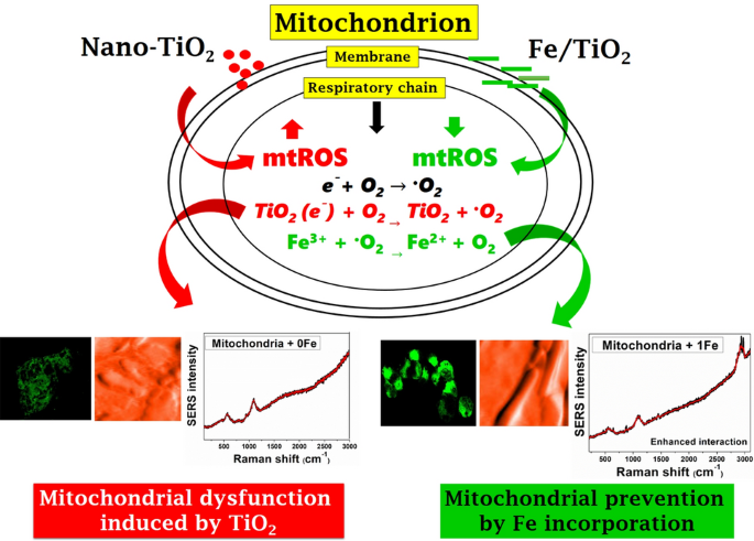
The paper assessed the toxic effect of titanium dioxide (TiO2) nanoparticles (NPs) on isolated mitochondria and its dysfunction prevention after Iron (Fe) incorporation. TiO2 and Fe content TiO2 NPs were synthesized and characterized using XPS, PL spectroscopy, and TEM. The nanostructure interaction with isolated mitochondria was investigated using circular dichroism (CD) confocal microscopy, flow cytometry, atomic force microscopy (AFM), surface-enhanced Raman spectroscopy (SERS), and FT-IR spectroscopy via nonspecific pathway. Fe content TiO2 NPs helps to control the dissolution rate of parent nanomaterial of TiO2 on the mitochondrial membrane. Confocal micrographs and flow cytometry results confirmed that Rhodamine 123 dye intensity get increased after interaction with Fe content TiO2 NPs which states the integrity of the mitochondrial membrane. AFM results revealed that TiO2 induces the swelling of mitochondrial tubules and also impaired the mitochondrial structure, whereas Fe content TiO2 NPs interaction prevents the impairment of mitochondrial tubules. The denaturation of a membrane protein by TiO2 interactions was observed through CD Spectroscopy. Further, nano-bio-interface study was performed using SERS, through shifting and extinct of peaks affiliated to membrane proteins and lipids. However, Fe content TiO2-treated samples showed a significant increase in the membrane potential of mitochondria via flow cytometry results.
Graphic Abstract
中文翻译:
TiO 2纳米颗粒诱导的线粒体功能异常的蛋白质和膜完整性研究及铁掺入的预防
本文评估了二氧化钛(TiO 2)纳米粒子(NPs)对孤立的线粒体的毒性作用以及铁(Fe)掺入后对功能障碍的预防作用。合成了TiO 2和Fe含量的TiO 2 NP,并使用XPS,PL光谱和TEM对其进行了表征。使用圆二色性(CD)共聚焦显微镜,流式细胞仪,原子力显微镜(AFM),表面增强拉曼光谱(SERS)和FT-IR光谱通过非特异性途径研究了与分离的线粒体的纳米结构相互作用。铁含量的TiO 2 NPs有助于控制TiO 2母体纳米材料的溶解速率在线粒体膜上。共焦显微照片和流式细胞仪结果证实,若丹明123与铁含量的TiO 2 NPs相互作用后,染料强度增加,表明线粒体膜的完整性。原子力显微镜的结果表明,TiO 2诱导线粒体小管肿胀,并损害线粒体结构,而Fe含量的TiO 2 NPs相互作用阻止了线粒体小管的损伤。通过CD光谱法观察到TiO 2相互作用引起的膜蛋白的变性。此外,通过转移和消除与膜蛋白和脂质相关的峰,使用SERS进行了纳米生物接口研究。但是,Fe含量为TiO 2流式细胞仪检测结果表明,经处理的样品线粒体膜电位显着增加。


 京公网安备 11010802027423号