Cardiovascular Pathology ( IF 3.7 ) Pub Date : 2021-03-21 , DOI: 10.1016/j.carpath.2021.107335 Georgia Karpathiou 1 , Jean Marc Dumollard 1 , Florian Camy 1 , Viviana Sramek 1 , Maroa Dridi 1 , Tiphanie Picot 1 , Mousa Mobarki 2 , Michel Peoc'h 1
|
Aims
Cardiac myxomas are rare tumors of incompletely elucidated pathogenesis. The aim of this study is to explore the possible presence of a senescence phenotype in cardiac myxomas, associated with an inflammatory and vasculogenic tumor microenvironment.
Methods and results
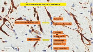
This is a retrospective study of 29 cardiac myxomas with immunohistochemical detection of various inflammatory, vascular, and senescence markers. We show that all myxomas contain tumor cells in senescence overexpressing p16, and a fraction of senescent endothelial cells. Macrophages are the principal inflammatory cell population, followed by cytotoxic T cells, with fewer plasma cells, mastocytes, and B lymphocytes. These populations are found in different intratumoral localizations. Larger tumor volume is associated with a lower percentage of myxoid matrix, higher cellularity, higher macrophage, and lower number of mast cells as well as higher PD-L1 expression by inflammatory cells. Higher vascular density is associated with higher percentage of B cells, a lower number of macrophages and higher number of mastocytes, and lower PD-L1 expression by inflammatory cells. Tumors with higher vascular density and higher cellularity show higher amounts of p16 senescent endothelial cells.
Conclusions
Myxoma tumor cells are in senescence and reside inside a tumor microenvironment with a distinct inflammatory profile rich in macrophages and cytotoxic T cells, and a rich vasculature, probably attributed to a senescence-associated secretory phenotype.
中文翻译:
心脏粘液瘤的衰老、免疫微环境和血管化
宗旨
心脏粘液瘤是发病机制不完全阐明的罕见肿瘤。本研究的目的是探索心脏粘液瘤中可能存在的衰老表型,这与炎症和血管源性肿瘤微环境有关。
方法和结果
这是一项对 29 例心脏粘液瘤的回顾性研究,免疫组化检测了各种炎症、血管和衰老标志物。我们发现所有粘液瘤都含有过表达 p16 的衰老肿瘤细胞和一部分衰老的内皮细胞。巨噬细胞是主要的炎症细胞群,其次是细胞毒性 T 细胞,浆细胞、肥大细胞和 B 淋巴细胞较少。这些群体存在于不同的肿瘤内定位中。较大的肿瘤体积与较低比例的粘液样基质、较高的细胞构成、较高的巨噬细胞和较低的肥大细胞数量以及炎症细胞较高的 PD-L1 表达相关。较高的血管密度与较高的 B 细胞百分比、较少数量的巨噬细胞和较多数量的肥大细胞有关,并降低炎症细胞的 PD-L1 表达。具有更高血管密度和更高细胞结构的肿瘤显示出更高数量的 p16 衰老内皮细胞。
结论
粘液瘤肿瘤细胞处于衰老状态,位于肿瘤微环境中,具有独特的炎症特征,富含巨噬细胞和细胞毒性 T 细胞,以及丰富的脉管系统,可能归因于衰老相关的分泌表型。


 京公网安备 11010802027423号